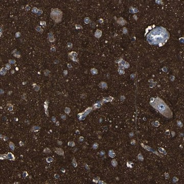
ABN1657
Anti-RME-8
from rabbit, purified by affinity chromatography
Synonym(s):
DnaJ homolog subfamily C member 13, Required for receptor-mediated endocytosis 8, RME-8
About This Item
WB
western blot: suitable
Recommended Products
biological source
rabbit
Quality Level
antibody form
affinity isolated antibody
antibody product type
primary antibodies
clone
polyclonal
purified by
affinity chromatography
species reactivity
human, monkey, rat, mouse
technique(s)
immunocytochemistry: suitable
western blot: suitable
NCBI accession no.
UniProt accession no.
shipped in
ambient
target post-translational modification
unmodified
Gene Information
human ... DNAJC13(23317)
General description
Specificity
Immunogen
Application
Western Blotting Analysis: A representative lot detected RME-8 associated with the 20,000 xg pelleted insoluble fraction from the HEPES buffer homogenates of human skin fibroblast MCH46, breast epithelial MCF10A, and embryonic kidney HEK293T cells (BT-474, HeLa, SK-BR-3) cells (Ioannou, M.S., et al. (2016). J. Biol. Chem. In press).
Western Blotting Analysis: A 1:1,000 dilution from a representative lot detected shRNA-mediated RME-8 downregulation in HEK293T cells (Courtesy of Peter McPherson, Ph.D., FRSC, and Martine Girard, PhD, McGill University, Canada).
Immunocytochemistry Analysis: A representative deteced a reduced RME-8 immunoreactivity in HeLa cells transfected with shRNA targeting either RME-8 or Spastin (Freeman, C.L., et al. (2014). J. Cell Sci. 127(Pt 9):2053-2070).
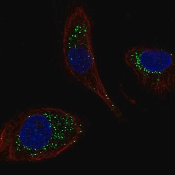
Immunocytochemistry Analysis: A representative lot detected puntate endosomal RME-8 immunoreactivity by fluorescent immunocytochemistry staining of 2% paraformaldehyde-fixed, 0.2% Triton X-100-permeabilized COS-7 cells. LIttle RME-8 immunoreactivity was seen associated with TGN, lysosome, or plasma membrane, RME-8 shRNA transfection greately abolished cellular immunostaining (Girard, M., et al. (2005). J. Biol. Chem. 280(48):40135-40143).
Western Blotting Analysis: A representative lot detected the presence of RME-8 in WASH complex immunoprecipitated from HeLa cells (BT-474, HeLa, SK-BR-3) cells (Freeman, C.L., et al. (2014). J. Cell Sci. 127(Pt 9):2053-2070).
Western Blotting Analysis: Representative lots detected a greatly reduced ~200 kDa target band in lysates from RME-8 shRNA-transfected monkey (COS-7) and human (BT-474, HeLa, SK-BR-3) cells (Freeman, C.L., et al. (2014). J. Cell Sci. 127(Pt 9):2053-2070; Girard, M., and McPherson, P.S. (2008). FEBS Lett. 582(6):961-966; Girard, M., et al. (2005). J. Biol. Chem. 280(48):40135-40143).
Western Blotting Analysis: A representative lot detected the ~200 kDa RME-8 target band in postnuclear extracts from multiple rat tissues as well as from human, monkey, mouse, and rat cell lines. RME-8 was found enriched in rat kidney tissue-derived subcellular fractions representing microsomes and clathrin-coated vesicles (CCVs), but not cytosol or plasma membrane (Girard, M., et al. (2005). J. Biol. Chem. 280(48):40135-40143).
Neuroscience
Quality
Western Blotting Analysis: A 1:500 dilution of this antibody detected RME-8 in 10 µg of NIH/3T3 cell lysate.
Target description
Physical form
Storage and Stability
Note: Variability in freezer temperatures below -20°C may cause glycerol containing solutions to become frozen during storage.
Other Notes
Disclaimer
Not finding the right product?
Try our Product Selector Tool.
Storage Class Code
10 - Combustible liquids
WGK
WGK 2
Certificates of Analysis (COA)
Search for Certificates of Analysis (COA) by entering the products Lot/Batch Number. Lot and Batch Numbers can be found on a product’s label following the words ‘Lot’ or ‘Batch’.
Already Own This Product?
Find documentation for the products that you have recently purchased in the Document Library.
Our team of scientists has experience in all areas of research including Life Science, Material Science, Chemical Synthesis, Chromatography, Analytical and many others.
Contact Technical Service


