immunohistochemistry (frozen sections): suitable microarray: suitable western blot: 1:500 using a fresh total rat brain extract or an enriched microtubule protein preparation
Monoclonal Anti-MAP1 (mouse IgG1 isotype) is derived from the HM-1 hybridoma produced by the fusion of mouse myeloma cells and splenocytes from BALB/c mice immunized with a rat brain MAPs preparation. MAP1 is one of the major neuronal MAPs as well as being the largest (350 kD). MAP1 is more generally distributed, being found in both dendrites and axons of neurons and in glial cells in brain, in chromatophores and on both interphase and mitotic microtubules in various tissue culture cells.
Specificity
The antibody does not cross-react with other MAPs or tubulin. By immunohistochemical staining of brain tissue, the antibody shows selective labeling of neurons with stronger staining of axons than dendrites.
Immunogen
rat brain microtubule-associated proteins (MAPs)
Application
Monoclonal Anti-MAP1 has been used in :
immunoblotting
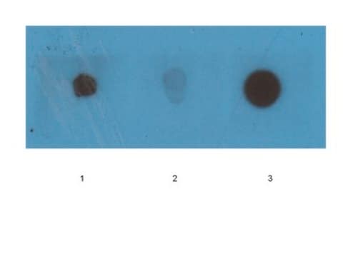
dot blot
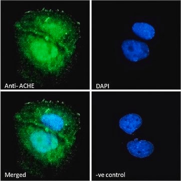
immunocytochemistry
immunohistochemistry
Biochem/physiol Actions
Microtubules are the ubiquitous cytoskeletal structural components that are involved in intracellular transport. They are composed of tubulin and microtubule-associated proteins (MAPs). MAPs are known to mediate the binding of membranous organelles, actin filaments and intermediate filaments to microtubules. Therefore, it might be important for cellular processes such as mitosis and organelle transport, and for determining the dynamic properties of the cytoskeleton.
Disclaimer
Unless otherwise stated in our catalog or other company documentation accompanying the product(s), our products are intended for research use only and are not to be used for any other purpose, which includes but is not limited to, unauthorized commercial uses, in vitro diagnostic uses, ex vivo or in vivo therapeutic uses or any type of consumption or application to humans or animals.
Molecular therapy : the journal of the American Society of Gene Therapy, 8(6), 886-894 (2003-12-11)
We have initiated studies to determine the feasibility of employing the Semliki Forest virus (SFV) expression system as a central nervous system (CNS) vector. We investigated the effects of infecting Balb/c mice intranasally (i.n.) with recombinant SFV particles expressing the
Effect of intranasal administration of semliki forest virus recombinant particles expressing reporter and cytokine genes on the progression of experimental autoimmune encephalomyelitis
Our team of scientists has experience in all areas of research including Life Science, Material Science, Chemical Synthesis, Chromatography, Analytical and many others.