Skip to Content
Merck
All Photos(1)

Documents

C8992

Sigma-Aldrich

Anti-phospho-Cofilin (pSer3) antibody produced in rabbit

IgG fraction of antiserum, buffered aqueous solution

Synonym(s):

Anti-18 kDa phosphoprotein, Anti-CFL1, Anti-Cofilin 1 (non-muscle), Anti-p18

Sign Into View Organizational & Contract Pricing


About This Item

MDL number:
UNSPSC Code:
12352203
NACRES:
NA.41

biological source

rabbit

Quality Level

conjugate

unconjugated

antibody form

IgG fraction of antiserum

antibody product type

primary antibodies

clone

polyclonal

form

buffered aqueous solution

mol wt

antigen ~19 kDa

species reactivity

mouse, human, pig (predicted), rat (predicted)

technique(s)

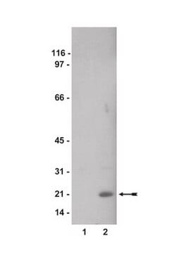
western blot: 1:2,000-1:4,000 using whole extracts of HeLa human epithelioid carcinoma and mouse NIH3T3 cells

UniProt accession no.

shipped in

dry ice

storage temp.

−20°C

target post-translational modification

phosphorylation (pSer3)

Gene Information

human ... CFL1(1072)
mouse ... Cfl1(12631)
rat ... Cfl1(29271)

Related Categories

General description

Cofilin is a small phosphoinositide-sensitive actin-binding protein capable of depolymerizing actin-filaments in vitro. It has gelsolin-like actin filament-severing protein similar to destrin/ADF (Actin Depolymerizing Factor). Cofilin has has a short β-strand at the C terminus. In mammals it has two isoforms: non-muscle (NM-CF, CF-L1) and muscle (M-CF, CF-L2). The protein is ubiquitiously present in tissues of eukaryotes and is especially abundant in neuronal tissues. It can shuttle between the cytoplasm and the nucleus in response to various stresses or signals, and may translocate from the cytoplasm to the plasma membrane in various cells.

Immunogen

synthetic phosphopeptide corresponding to amino acids 2-9 (pSer3) of human cofilin with a C-terminal added cysteine, conjugated to KLH. This sequence is identical in mouse, rat, and pig.

Application

Anti-phospho-Cofilin antibody was used:
  • as primary antibody for immunoblottingto to study the role of the Rac1 GTPase.
  • for western blot analysis in a comparative study of signals induced by integrin ligation during cell attachment, mechanical force from intracellular contraction, or cell stretching by external force.
  • as primary antibody for western blotting in a study to detect the level of cofilin.

Biochem/physiol Actions

Cofilin binds stoichiometrically to monomeric G-actin and actin protomers in filaments in an apparently pH-dependent, Ca2+ independent manner. Cofilin intercalates between longitudinally associated actin monomers within the filament and distorts its helical twist.It cleaves the filaments and accelerates actin subunits dissociation from their ‘pointed′ ends under specific conditions. Cofilin is essential for viability and is important for many cellular processes involving actin remodeling such as motility at the leading edge of cells, polarized cell growth, endocytosis, phagocytosis, cellular activation, cytokinesis, and pathogen intracellular motility. It′s activity is regulated through reversible phosphorylation and dephosphorylation. In phosphorylated form, it behaves inactive and unable to bond with actin. Phosphorylation of cofilin is regulated in vertebrates by at least four protein kinases: LIM Kinase 1, LIM Kinase 2, Testicular Kinase 1, and Testicular Kinase 2. The dephosphorylation of cofilin enables its actin severing and depolymerizing activity and drives directional cell motility, thus providing a simple phosphoregulatory mechanism for actin reorganization.

Physical form

Solution in 0.01 M phosphate buffered saline, pH 7.4, containing 15 mM sodium azide.

Disclaimer

Unless otherwise stated in our catalog or other company documentation accompanying the product(s), our products are intended for research use only and are not to be used for any other purpose, which includes but is not limited to, unauthorized commercial uses, in vitro diagnostic uses, ex vivo or in vivo therapeutic uses or any type of consumption or application to humans or animals.

Not finding the right product?  

Try our Product Selector Tool.

Storage Class Code

10 - Combustible liquids

WGK

WGK 3

Flash Point(F)

Not applicable

Flash Point(C)

Not applicable

Personal Protective Equipment

dust mask type N95 (US), Eyeshields, Gloves

Certificates of Analysis (COA)

Search for Certificates of Analysis (COA) by entering the products Lot/Batch Number. Lot and Batch Numbers can be found on a product’s label following the words ‘Lot’ or ‘Batch’.

Already Own This Product?

Find documentation for the products that you have recently purchased in the Document Library.

Visit the Document Library

Christopher A Foote et al.
American journal of physiology. Heart and circulatory physiology, 310(2), H188-H198 (2015-11-15)
Inward remodeling of the resistance vasculature is strongly associated with life-threatening cardiovascular events. Previous studies have demonstrated that both actin polymerization and the activation of transglutaminases mediate early stages of the transition from a structurally normal vessel to an inwardly
S Arber et al.
Nature, 393(6687), 805-809 (1998-07-09)
Cell division, cell motility and the formation and maintenance of specialized structures in differentiated cells depend directly on the regulated dynamics of the actin cytoskeleton. To understand the mechanisms of these basic cellular processes, the signalling pathways that link external
Kateřina Kuželová et al.
PloS one, 9(3), e92560-e92560 (2014-03-26)
P21-activated kinases (PAKs) are involved in the regulation of multiple processes including cell proliferation, adhesion and migration. However, the current knowledge about their function is mainly based on results obtained in adherent cell types. We investigated the effect of group
J R Bamburg
Annual review of cell and developmental biology, 15, 185-230 (1999-12-28)
Ubiquitous among eukaryotes, the ADF/cofilins are essential proteins responsible for the high turnover rates of actin filaments in vivo. In vertebrates, ADF and cofilin are products of different genes. Both bind to F-actin cooperatively and induce a twist in the
Kathrin S Zeller et al.
PloS one, 8(5), e64897-e64897 (2013-06-06)
Cells are exposed to several types of integrin stimuli, which generate responses generally referred to as "integrin signals", but the specific responses to different integrin stimuli are poorly defined. In this study, signals induced by integrin ligation during cell attachment

Our team of scientists has experience in all areas of research including Life Science, Material Science, Chemical Synthesis, Chromatography, Analytical and many others.

Contact Technical Service