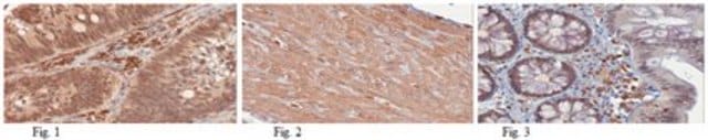
immunohistochemistry (formalin-fixed, paraffin-embedded sections): 1:100 using sections of human colon carcinoma microarray: suitable western blot: 1:2,000 using human epidermal carcinoma A431 whole cell extract
Bak (Bcl-2 homologous antagonist/killer, Bak1) belongs to the B-cell lymphoma 2 (Bcl-2) family of proteins. The bak gene is mapped to chromosome 6 and encodes a 233 amino acid protein with a predicted MW of 23.4 kDa. Bak shares homology with Bcl-2 in the Bcl‐2 homology (BH) domains (BH1 and BH2). Bak is expressed in a wide variety of cell types and tissues, with the highest levels observed in heart and skeletal muscle.
Immunogen
synthetic peptide corresponding to the N-terminus of human Bak amino acids 23-38 with C-terminally added lysine, conjugated to KLH.
Application
Anti-Bak antibody produced in rabbit has been used in immunoblotting and immunohistochemistry.[1]
Biochem/physiol Actions
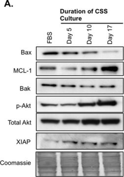
Bak (Bcl-2 homologous antagonist/killer, Bak1) is involved in regulating apoptosis. Bak can accelerate the rate of apoptosis when overexpressed in some cell lines. Increased Bak expression in normal and neoplastic intestinal epithelial cells results in apoptosis. However, expression of Bak in a human lymphoblastoid cell line, provided protection from apoptosis induced by serum deprivation and the oxidant menadione, suggesting that the function of Bak may be context dependent.
Physical form
Solution in 0.01 M phosphate buffered saline, pH 7.4, containing 15 mM sodium azide.
Disclaimer
Unless otherwise stated in our catalog or other company documentation accompanying the product(s), our products are intended for research use only and are not to be used for any other purpose, which includes but is not limited to, unauthorized commercial uses, in vitro diagnostic uses, ex vivo or in vivo therapeutic uses or any type of consumption or application to humans or animals.
The Journal of biological chemistry, 288(36), 26027-26038 (2013-07-31)
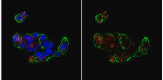
Bak and Bax are the essential effectors of the intrinsic pathway of apoptosis. Following an apoptotic stimulus, both undergo significant changes in conformation that facilitates their self-association to form pores in the mitochondrial outer membrane. However, the molecular structures of
Association of Bax and Bak Homo-oligomers in Mitochondria Bax REQUIREMENT FOR Bak REORGANIZATION AND CYTOCHROMEc RELEASE
During apoptosis, Bak and Bax undergo major conformational change and form symmetric dimers that coalesce to perforate the mitochondrial outer membrane via an unknown mechanism. We have employed cysteine labelling and linkage analysis to the full length of Bak in
DNA damage-related gene expression as biomarkers to assess cellular response after gamma irradiation of a human lymphoblastoid cell line
Our team of scientists has experience in all areas of research including Life Science, Material Science, Chemical Synthesis, Chromatography, Analytical and many others.