Skip to Content
MilliporeSigma
All Photos(1)

Key Documents

MAB3060

Sigma-Aldrich

Anti-Paxillin Antibody, clone 349

clone 349, Chemicon®, from mouse

Synonym(s):

Anti-AW108311, Anti-AW123232, Anti-Pax

Sign Into View Organizational & Contract Pricing


About This Item

UNSPSC Code:
12352203
eCl@ss:
32160702
NACRES:
NA.41

biological source

mouse

Quality Level

antibody form

purified immunoglobulin

antibody product type

primary antibodies

clone

349, monoclonal

species reactivity

rat, human, mouse, canine, chicken

manufacturer/tradename

Chemicon®

technique(s)

ELISA: suitable
immunocytochemistry: suitable
immunohistochemistry: suitable
western blot: suitable

isotype

IgG1

NCBI accession no.

UniProt accession no.

shipped in

wet ice

target post-translational modification

unmodified

Gene Information

human ... PXN(5829)
rat ... Pxn(360820)

Specificity

Reacts strongly with two bands on Western blots of total cell lysate from chick embryo fibroblasts or rat brain. The lower molecular band is believed to be either an isoform or a degradation product of the 68 kD protein, since it is found also on SDS-PAGE of affinity-purified paxillin.

Immunogen

Affinity purified phosphotyrosine-containing proteins from Rous sarcoma virus-transformed chick embryo fibroblasts.

Application

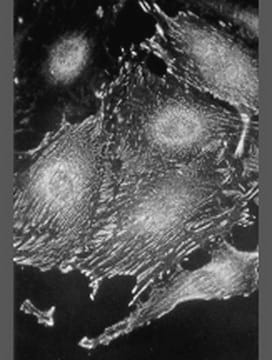
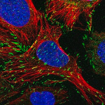
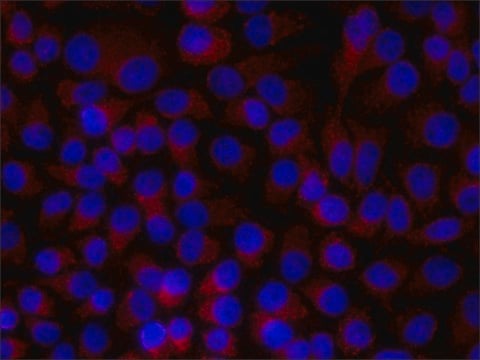
Anti-Paxillin Antibody, clone 349 detects level of Paxillin & has been published & validated for use in ELISA, WB, IC, IH.
Research Category
Signaling
Research Sub Category
Cytoskeletal Signaling
Western blot: 1:1,000-1:10,000

ELISA

Immunohistochemistry 1:900 in Placenta

Immunocytochemistry

Immunoprecipitation

Optimal working dilutions must be determined by end user.

Linkage

Replaces: 04-581

Physical form

Format: Purified
Purified immunoglobulin. Liquid in aqueous buffered solution containing 0.09% sodium azide.

Storage and Stability

Maintain at -20°C in undiluted aliquots for up to 12 months after date of receipt. Avoid repeated freeze/thaw cycles.

During shipment, small volumes of product will occasionally become entrapped in the seal of the product vial. For products with volumes of 200 μL or less, we recommend gently tapping the vial on a hard surface or briefly centrifuging the vial in a tabletop centrifuge to dislodge any liquid in the container′s cap.

Analysis Note

Control
POSITIVE CONTROL:

For western blot, load 5-10 μg of whole cell lysate prepared from the human epidermoid carcinoma cell line, A431.

Other Notes

Concentration: Please refer to the Certificate of Analysis for the lot-specific concentration.

Legal Information

CHEMICON is a registered trademark of Merck KGaA, Darmstadt, Germany

Disclaimer

Unless otherwise stated in our catalog or other company documentation accompanying the product(s), our products are intended for research use only and are not to be used for any other purpose, which includes but is not limited to, unauthorized commercial uses, in vitro diagnostic uses, ex vivo or in vivo therapeutic uses or any type of consumption or application to humans or animals.

Not finding the right product?  

Try our Product Selector Tool.

Storage Class Code

12 - Non Combustible Liquids

WGK

WGK 1


Certificates of Analysis (COA)

Search for Certificates of Analysis (COA) by entering the products Lot/Batch Number. Lot and Batch Numbers can be found on a product’s label following the words ‘Lot’ or ‘Batch’.

Already Own This Product?

Find documentation for the products that you have recently purchased in the Document Library.

Visit the Document Library

Role of zyxin in differential cell spreading and proliferation of melanoma cells and melanocytes.
van der Gaag, EJ; Leccia, MT; Dekker, SK; Jalbert, NL; Amodeo, DM; Byers, HR
The Journal of Investigative Dermatology null
M Ishino et al.
FEBS letters, 474(2-3), 179-183 (2000-06-06)
Hic-5 is a CAKbeta-binding protein localized at focal adhesions. Here we show that overexpression of CAKbeta or Fyn, but not FAK, enhanced the tyrosine phosphorylation of coexpressed Hic-5 in COS-7 cells. These phosphorylations were further augmented by stimulating cells with
Imaging morphogenesis, in Xenopus with Quantum Dot nanocrystals.
Stylianou, P; Skourides, PA
Mechanisms of Development null
Mariam Mansour et al.
Journal of cell science, 129(2), 277-289 (2015-12-09)
Eph receptors and their corresponding membrane-bound ephrin ligands regulate cell positioning and establish tissue patterns during embryonic and oncogenic development. Emerging evidence suggests that assembly of polymeric Eph signalling clusters relies on cytoskeletal reorganisation and underlies regulation by protein tyrosine

Our team of scientists has experience in all areas of research including Life Science, Material Science, Chemical Synthesis, Chromatography, Analytical and many others.

Contact Technical Service