Saltar al contenido
Merck
Home3D Cell CultureBarrier Formation and Permeability Assays using Millicell® Hanging Cell Culture Inserts

Barrier Formation and Permeability Assays using Millicell® Hanging Cell Culture Inserts

Barrier Assay Overview

Cell culture barrier assays are used to model barrier tissues (kidney, lung, intestinal, etc.) in drug delivery, drug absorbance, wound healing, bacterial/viral infections, inflammation and other studies. These barriers can be formed in vitro when epithelial cells are seeded as a single cell suspension and grow together until fused into one tissue. Typically, this tissue will be one epithelial cell in thickness and is termed the epithelial monolayer. In standard barrier assays, cells are seeded onto porous membranes and observed noninvasively via trans-epithelial electrical resistance (TEER) measurements. TEER measurements are validated, label-free and fast techniques for indicating epithelial monolayer barrier formation before further evaluation and experimentation. Barrier formation is observed by the increase in resistance measure; a confluent epithelial monolayer will plateau at higher resistance values. For most barrier assays, an epithelial monolayer is the desired type of cell barrier because it models the single-cell thickness of lung-to-blood, kidney filtration, or intestinal absorption and can be used to analyze the paracellular space between epithelial cells.  Once the integrity of the cellular barrier has been confirmed, these tissues can then be used in experiments that probe barrier permeability, disruption, or strength.

Tight junction pores are physiological gatekeepers of paracellular transport in epithelial tissues. Paracellular permeability assays assess monolayer barrier formation and tightness of paracellular tight junctions by measuring the amount of a molecule that passes through an epithelial cell monolayer in a given amount of time. This type of assay requires live cells and is usually non-destructive (e.g., Lucifer yellow paracellular permeability assay). Immunostaining and tissue immunocytochemistry (ICC) are commonly used for visualization, pre-empting the need for electron microscopy. Many of these analyses are focused on barrier pass-through tracked as dye, drug conjugates, or amount of target protein transferred. Alternative analyses involve visualizing the membrane using stains such as crystal violet, fluorescent antibody staining, and ICC.
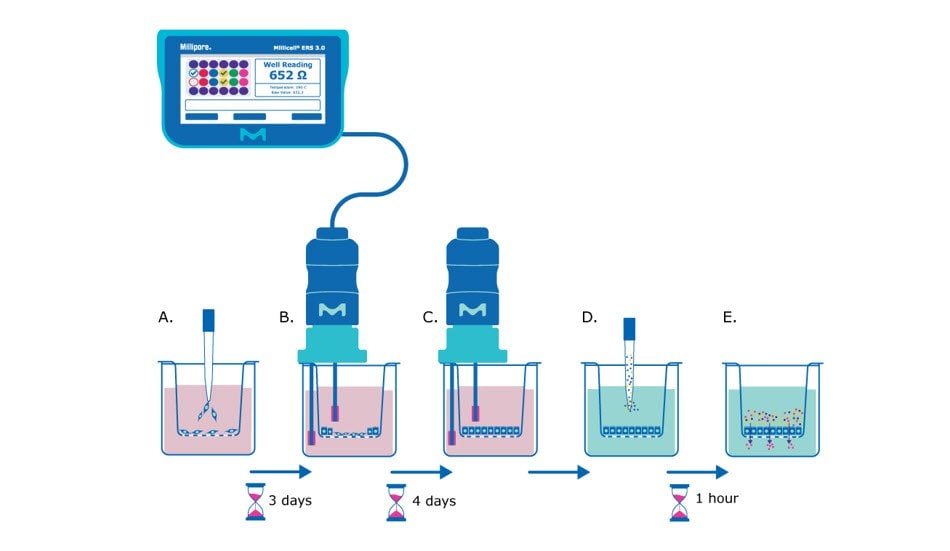
Barrier assay principle using Millicell® hanging cell culture inserts. The cells are seeded in the insert and after three days researchers use the Millicell 3.0 ERS to take TEER measurements. 7 days after seeding the TEER measurements should plateau, the media can be exchanged, and a permeability assay dye can be added. Small molecules can be added and incubated with the monolayer and pass-through can be measured.

Figure 1. Barrier assay principle using Millicell® hanging cell culture inserts. A) On day 0, epithelial cells are seeded in single-cell suspension onto the Millicell® hanging cell culture insert membrane. B) After several days (~3 days after seeding for MDCK cells) the cells will be confluent enough to begin taking TEER measurements, assessing the epithelial cell monolayer formation based on the resistance value. C) TEER measurements can be taken daily or every other day to track monolayer barrier formation until cells are seen to plateau at a consistent resistance value (~7 days after seeding for MDCK cells). D) At this point the media is exchanged for barrier assay liquid (Hank’s balanced salt solution) and permeability assay dye/molecule (e.g., Lucifer yellow dye) is added to the apical side of the membrane. E) The small molecules are incubated with the epithelial monolayer at 37°C for recommended protocol length (e.g., 1 hour), and then the liquid from the basolateral side of the membrane insert is evaluated for the amount of pass-through of the chosen molecule. 

What do I need to run a Lucifer yellow barrier assay?

  • Millicell® hanging cell culture insert with 0.4 or 1 µm pore size (see Selecting a Millicell® Hanging Cell Culture Insert below)

Selecting a Millicell® hanging cell culture insert

Barrier assays are performed on hanging inserts as porous membranes offer vastly improved characteristics over standard polystyrene surfaces for what is called 2.5D cell culturing. Membranes in these inserts differ in terms of pore size, which becomes important for different assay types. For example, migration assays will have the widest range of pore sizes based on the cell type involved in the experiment.

Table 1.Millicell® cell culture insert membrane pore size and companion assays.

TEER Detection Method

The Millicell® ERS 3.0 voltohmmeter uses low current and voltages to allow for non-destructive testing for epithelial monolayer confluence in cell cultures. TEER instruments produce a low AC current that avoids electrode metal deposits and adverse effects on tissues which can otherwise be caused by higher DC currents. Confluence of a cellular monolayer is determined by an increase or a plateau in tissue resistance detected using TEER readers.

MDCK culture media

Seeding cells onto a 24-well Millicell® hanging cell culture insert

  1. Culture MDCK cells until they are 60-80% confluent.
  2. Aspirate media from the culture flask.
  3. Add sterile PBS for 3 minutes at room temperature.
  4. Aspirate liquid from the culture flask.
  5. Add trypsin to the culture flask and incubate in a 37°C, 5% CO2 cell culture incubator.
  6. Stop the trypsin reaction by adding equal parts complete cell culture media to the flask.
  7. Collect liquid suspension and spin down in the centrifuge at 300 x g for 3 minutes.
  8. Resuspend cells in 1 mL of cell complete culture media (see MDCK culture media recipe above).
  9. Count cells using trypan blue and hemocytometer. Mix 90% trypan blue with 10% cell suspension (e.g., 180 µL trypan blue and 20 µL cell suspension).
  10. Count live cells on the hemocytometer and calculate cells per milliliter.
  11. Add 900 µL of complete media to the basolateral side of the 24-well membranes.
  12. Resuspend cell suspension in appropriate volumes to dispense 200 µL of suspension containing the desired cell seeding density (e.g., 1,250 cells/insert, 2,500 cells/insert, 5,000 cells/insert).
  13. Place in the 37°C, 5% carbon dioxide cell culture incubator and change media every other day.
    NOTE: Best practice is to pre-warm complete cell culture media before adding to Millicell® hanging cell culture insert and receiver plate.

Measuring cell resistance

  1. Allow plate reach to reach room temperature (15-30 minutes).
  2. Load plate map and assign wells to groups on the Millicell® ERS 3.0 Digital Voltohmmeter (see p. 11 of user guide).
  3. Test the Millicell® ERS 3.0 Digital Voltohmmeter using the verification adapter (see p. 22 of user guide).
  4. Set the MODE switch to Ohms and turn the Power switch On.
  5. Immerse the electrode so that the shorter tip is in the apical side of the Millicell® hanging cell culture insert, and the longer tip is in the outer well. The shorter tip should not contact cells growing on the membrane and the longer tip should just touch the bottom of the outer well. To ensure stable and reproducible results, make sure that the electrode is held steady at a 90° angle to the plate and insert bottom.
    NOTE: If rinsing is required between measurements to prevent sample carryover, use cell culture media rather than distilled water.
  6. Record the resistance.
  7. Determine blank resistance by adding media or electrolyte solution to a blank well/cell culture insert (i.e., the cell culture insert without cells and complete cell culture media).
  8. Measure the resistance across the blank insert and use this value as a “zero” or “background” resistance level. To obtain true tissue resistance, subtract the resistance reading across the blank insert from the resistance reading across the tissue (cell culture insert with cells).
  9. The resistance is inversely proportional to the area of the tissue. The larger the membrane, the lower the resistance.
    Unit Area Resistance (Ω × cm2) = Resistance (Ω) × Effective membrane Area (cm2)
    NOTE: Resistance can vary based on sample temperature, pH, and electrode depth in solution.

TEER Application Notes, Guides, and Other Resources

Lucifer Yellow Barrier Assay Sample Protocol

Steps

  1. Aspirate media from the apical and basolateral sides of the membrane/well.
    TIP: Be careful not to touch or poke the membrane while aspirating.
  2. Wash/rinse membrane and well with prewarmed Hank’s Balanced Salt Solution (HBSS).
    TIP: Prewarmed HBSS has the least impact on the cell monolayer; the use of PBS will be more harmful and cause a greater percentage of dye passthrough.
  3. Aspirate HBSS from the apical and basolateral sides of the membrane/well.
    TIP: Be careful not to touch or poke the membrane while aspirating.
  4. Dispense 900 µL HBSS to the basolateral side of the membrane.
  5. Dispense 200 µL of Lucifer yellow dye (100 µg/mL) to the apical side of the membrane.
    TIP: Be very careful not to accidentally add Lucifer yellow dye or spill/drip into the basolateral compartment. This will disrupt assay result accuracy. Do not poke the membrane when pipetting.
  6. Place in 37°C, 5% CO2 incubator for 1 hour. Increase incubation time to 2-3 hours for low porosity membranes like the 0.4 µm clear Millicell® inserts (PCHT24H48). 
    TIP: For best results keep plate flat and do not shake.
  7. Use this time to make standards for the assay plate, which identify your sample values.
        a.  Make a 100% standard [Std(100)] by combining:
             200 µL of Lucifer yellow Dye (100 µg/mL) with 900 µL of HBSS.
        b.  Dilute standards 1:1 in a serial dilution.
             500 µL of Std(100) with 500 µL of HBSS would make the Std(50)
        c.  Lowest standard (besides 100% HBSS) should contain 0.39% Lucifer yellow (this would make nine standards and a tenth for HBSS
             "zero").
        d.  Add 100 µL of each standard to a fluorescence spectroscopy plate (in triplicate).
  8. Remove plate from incubator and collect 100 µL of fluid from the basolateral side of the membranes to place into the fluorescence spectroscopy plate.
    TIP: For best results, do this in triplicate so that results can be averaged as technical replicates.
  9. Read fluorescence spectroscopy plate on a plate reader and determine the fluorescent absorbance using 485 nm excitation and 535 nm emission wavelengths.
  10. Use the generated standard curve to calculate the Lucifer yellow concentration in each well. These values can then be used to determine the percent pass-through for each sample.
    RFU = relative fluorescence units assigned by the plate reader.
Formulas for RFU Equilibrium
  1. Typically, the success criterion for a Lucifer yellow permeability assay is set at less than five percent passthrough.

Sample Barrier Assay Data

MDCK barrier formation assay using Millicell® hanging cell culture inserts

Figure 3. All membranes depicted are high-density (1×10⁸) 0.4 µm pore size PET 24-well membranes. These results compare Millicell® hanging cell culture inserts (PTHT24H48) to two comparable brands of inserts. MDCK cells were seeded at the same seeding density in parallel across all three manufacturers. The three brands of inserts are not significantly different from each other on day 7 after seeding and follow a similar kinetic for the formation of a monolayer.

Lucifer yellow permeability assay using Millicell® hanging cell culture inserts

Figure 4. Lucifer yellow permeability assays test the formation of the cellular monolayer and tight junctions between cells. All membranes depicted are high-density (1 x 10⁸ pores) 0.4 µm pore size PET membranes in 24-well formats. These results compare the Millicell® hanging cell culture insert (PTHT24H48) to two other brands of inserts. This assay was performed on Day 7 of the assay in Figure 3. Barrier formation is deemed successful if there is less than 5% pass-through of the dye from the apical side of the membrane into the basolateral side of the membrane (pass-through depicted in the final panel in Figure 1). There were no statistical differences between these inserts.

TEER measurement of barrier integrity using a Millicell® ERS-2 voltohmmeter

Figure 5. TEER values are dependent on starting seeding densities, which correlate with the confluence of cells on the Millicell® hanging cell culture insert. Higher cell seeding densities will form a cellular monolayer faster. Depending on the experiment, it might be more beneficial to have higher or lower seeding densities depending on the study’s focus. In the example above, MDCK cells were seeded from the same stock and were measured for resistance on the days indicated. All measurements were taken on the Millicell® ERS 3.0 digital voltohmmeter with Millicell® hanging cell culture inserts (PTHT24H48). 

Barrier Assay Tips and Tricks

  • Choose a membrane that best fits your cell type and assay (see Table 1 above)
  • Change media every other day to ensure cells have enough nutrients and maintain vital cell signaling for monolayer/tight junction formation.
  • Do not leave cells outside of the incubator for prolonged periods of time before assays. This can negatively impact Lucifer yellow permeability assay results as well as other similar assays.
  • Make sure the electrodes on the TEER probe are fully submerged in media for the most stable readings.
  • Hold the TEER probe at the same depth in each well/insert for consistent and comparable readings.
  • To ensure stable and reproducible results, make sure that the electrode is positioned at a 90° angle to the plate insert.
  • Resistance can vary with temperature. TEER readings increase (higher ohms Ω) at lower temperatures (lower °C). Be sure to wait for samples to acclimate to room temperature before taking readings.
  • Resistance is affected by media pH. Higher carbon dioxide content will be present in samples that are pulled immediately out of an incubator and is associated with lower resistance values. The amount of carbon dioxide will equilibrate with the atmospheric carbon dioxide while acclimating to room temperature.
  • Clean TEER electrodes periodically with an enzymatic detergent such as Tergazyme® to eliminate protein buildup on the electrode from cell culture media. This cleaning procedure can be found in the Millicell® ERS 3.0 user guide.
  • The Millicell® ERS 3.0 model has a cable that is more vulnerable to interference from radio waves. These come from cell phones, laptops, computers, radiation equipment (MRI, x-ray, etc.), and even smart watches. Consider moving testing area to an area with the least amount of these disturbances.
  • Never put the Millicell® ERS 3.0 voltohmmeter or its electrode probe under UV light. This will damage the electrode sensor and lead to inaccurate, variable, or failure to take readings.
  • If rinsing is required between measurements to prevent sample carryover, use cell culture media rather than distilled water.
  • Be careful not to touch or poke the membrane while aspirating.
  • During a Lucifer yellow permeability assay, prewarmed HBSS has the least impact on the cell monolayer; the use of PBS will be more harmful and cause a greater percentage of dye pass-through.
  • Be very careful not to accidentally add Lucifer yellow dye or spill/drip into the basolateral compartment. This will disrupt assay result accuracy. Do not poke the membrane when pipetting.
  • Once Lucifer yellow dye is added to the membrane, for best results keep plate flat and do not shake.
  • At the end of the Lucifer yellow permeability assay, collect basolateral liquid after mixing (pipette up and down), then run passthrough results in technical triplicates for the best outcome.
Related Products
Loading

References

1.
Sawamiphak S, Ritter M, Acker-Palmer A. 2010. Preparation of retinal explant cultures to study ex vivo tip endothelial cell responses. Nat Protoc. 5(10):1659-1665. https://doi.org/10.1038/nprot.2010.130
2.
Tamari T, Kawar-Jaraisy R, Doppelt O, Giladi B, Sabbah N, Zigdon-Giladi H. The Paracrine Role of Endothelial Cells in Bone Formation via CXCR4/SDF-1 Pathway. Cells. 9(6):1325. https://doi.org/10.3390/cells9061325
3.
Hamdollah Zadeh M, Glass CA, Magnussen A, Hancox JC, Bates DO. 2008. VEGF-Mediated Elevated Intracellular Calcium and Angiogenesis in Human Microvascular Endothelial CellsIn Vitroare Inhibited by Dominant Negative TRPC6. Microcirculation. 15(7):605-614. https://doi.org/10.1080/10739680802220323
4.
Hadjichristou C, Papachristou E, Bonovolias I, Bakopoulou A. 2020. Three-dimensional tissue engineering-based Dentin/Pulp tissue analogue as advanced biocompatibility evaluation tool of dental restorative materials. Dental Materials. 36(2):229-248. https://doi.org/10.1016/j.dental.2019.11.013
5.
DU H, SHI H, CHEN D, ZHOU Y, CHE G. 2015. Cross-talk between endothelial and tumor cells via basic fibroblast growth factor and vascular endothelial growth factor signaling promotes lung cancer growth and angiogenesis. 9(3):1089-1094. https://doi.org/10.3892/ol.2015.2881
6.
Del Tatto M, Ng T, Aliotta JM, Colvin GA, Dooner MS, Berz D, Dooner GJ, Papa EF, Hixson DC, Ramratnam B, et al. 2011. Marrow cell genetic phenotype change induced by human lung cancer cells. Experimental Hematology. 39(11):1072-1080. https://doi.org/10.1016/j.exphem.2011.08.008
7.
Hoeks C, Vanheusden M, Peeters LM, Stinissen P, Broux B, Hellings N. Treg-Resistant Cytotoxic CD4+ T Cells Dictate T Helper Cells in Their Vicinity: TH17 Skewing and Modulation of Proliferation. IJMS. 22(11):5660. https://doi.org/10.3390/ijms22115660
8.
Sygnecka K, Heider A, Scherf N, Alt R, Franke H, Heine C. 2015. Mesenchymal Stem Cells Support Neuronal Fiber Growth in an Organotypic Brain Slice Co-Culture Model. Stem Cells and Development. 24(7):824-835. https://doi.org/10.1089/scd.2014.0262
9.
Maeda T, Ibi M, Shimazu S, Akaike A. 1998. Co-culture With the Striatum Attenuates N-Methyl-D-aspartate Cytotoxicity in Dopaminergic Neurons of Rat Mesencephalic Slice Cultures. Japanese Journal of Pharmacology. 77(2):161-168. https://doi.org/10.1254/jjp.77.161
10.
Whitman MC, Bell JL, Nguyen EH, Engle EC. Ex Vivo Oculomotor Slice Culture from Embryonic GFP-Expressing Mice for Time-Lapse Imaging of Oculomotor Nerve Outgrowth. JoVE.(149): https://doi.org/10.3791/59911
11.
Robinson J, Turkington S, Abey S, Kenea N, Henderson W. 2019. Differential gene expression and gene-set enrichment analysis in Caco-2 monolayers during a 30-day timeline with Dexamethasone exposure. Tissue Barriers. 7(3):e1651597. https://doi.org/10.1080/21688370.2019.1651597
12.
Ricciardolo FL, Folkerts G, Folino A, Mognetti B. 2018. Bradykinin in asthma: Modulation of airway inflammation and remodelling. European Journal of Pharmacology. 827181-188. https://doi.org/10.1016/j.ejphar.2018.03.017
13.
Vis MAM, Ito K, Hofmann S. Impact of Culture Medium on Cellular Interactions in in vitro Co-culture Systems. Front. Bioeng. Biotechnol.. 8 https://doi.org/10.3389/fbioe.2020.00911
14.
Matsushita T, Kibayashi T, Katayama T, Yamashita Y, Suzuki S, Kawamata J, Honmou O, Minami M, Shimohama S. 2011. Mesenchymal stem cells transmigrate across brain microvascular endothelial cell monolayers through transiently formed inter-endothelial gaps. Neuroscience Letters. 502(1):41-45. https://doi.org/10.1016/j.neulet.2011.07.021
15.
Kidney JC, Proud D. 2000. Neutrophil Transmigration across Human Airway Epithelial Monolayers. Am J Respir Cell Mol Biol. 23(3):389-395. https://doi.org/10.1165/ajrcmb.23.3.4068
16.
Van Oostveldt K, Paape M, Burvenich C. 2002. Apoptosis of Bovine Neutrophils Following Diapedesis Through a Monolayer of Endothelial and Mammary Epithelial Cells. Journal of Dairy Science. 85(1):139-147. https://doi.org/10.3168/jds.s0022-0302(02)74062-9
17.
Heise R, Vetter-Kauczok CS, Skazik C, Czaja K, Marquardt Y, Lue H, Merk HF, Bernhagen J, Baron JM. 2012. Expression and Function of Macrophage Migration Inhibitory Factor in the Pathogenesis of UV-Induced Cutaneous Nonmelanoma Skin Cancer?. 88(5):1157-1164. https://doi.org/10.1111/j.1751-1097.2012.01108.x
18.
Yu J, Kim HM, Kim KP, Son Y, Kim M, Park K. 2019. Ceramide kinase regulates the migration of bone marrow-derived mesenchymal stem cells. Biochemical and Biophysical Research Communications. 508(2):361-367. https://doi.org/10.1016/j.bbrc.2018.11.154
19.
Wang X, Ma G, Zhang R, Liu L, Zhu J, Zhu H. 2019. Molecular characterization and biological functioning of interleukin-8 in Siberian sturgeon (Acipenser baeri). Fish & Shellfish Immunology. 9091-101. https://doi.org/10.1016/j.fsi.2019.04.010
20.
Trost B, Walker S, Wang Z, Thiruvahindrapuram B, MacDonald JR, Sung WW, Pereira SL, Whitney J, Chan AJ, Pellecchia G, et al. 2018. A Comprehensive Workflow for Read Depth-Based Identification of Copy-Number Variation from Whole-Genome Sequence Data. The American Journal of Human Genetics. 102(1):142-155. https://doi.org/10.1016/j.ajhg.2017.12.007
21.
Spary LK, Salimu J, Webber JP, Clayton A, Mason MD, Tabi Z. 2014. Tumor stroma-derived factors skew monocyte to dendritic cell differentiation toward a suppressive CD14+PD-L1+phenotype in prostate cancer. OncoImmunology. 3(9):e955331. https://doi.org/10.4161/21624011.2014.955331
22.
Welch HC, Condliffe AM, Milne LJ, Ferguson GJ, Hill K, Webb LM, Okkenhaug K, Coadwell WJ, Andrews SR, Thelen M, et al. 2005. P-Rex1 Regulates Neutrophil Function. Current Biology. 15(20):1867-1873. https://doi.org/10.1016/j.cub.2005.09.050
23.
Zhang Y, Wang B, Meng X, Sun G, Gao C. 2011. Influences of Acid-Treated Multiwalled Carbon Nanotubes on Fibroblasts: Proliferation, Adhesion, Migration, and Wound Healing. Ann Biomed Eng. 39(1):414-426. https://doi.org/10.1007/s10439-010-0151-y
24.
Sokolowski JD, Chabanon-Hicks CN, Han CZ, Heffron DS, Mandell JW. Fractalkine is a â??find-meâ? signal released by neurons undergoing ethanol-induced apoptosis. Front. Cell. Neurosci.. 8 https://doi.org/10.3389/fncel.2014.00360
25.
Hagan K, Varelas P, Zheng H. 2022. Endocannabinoid System of the Blood?Brain Barrier: Current Understandings and Therapeutic Potentials. Cannabis and Cannabinoid Research. 7(5):561-568. https://doi.org/10.1089/can.2021.0101
26.
Tucci M, Ciavarella S, Strippoli S, Brunetti O, Dammacco F, Silvestris F. 2011. Immature dendritic cells from patients with multiple myeloma are prone to osteoclast differentiation in vitro. Experimental Hematology. 39(7):773-783.e1. https://doi.org/10.1016/j.exphem.2011.04.006
27.
Fell LH, Seiler-Mußler S, Sellier AB, Rotter B, Winter P, Sester M, Fliser D, Heine GH, Zawada AM. 2016. Impact of individual intravenous iron preparations on the differentiation of monocytes towards macrophages and dendritic cells. Nephrol. Dial. Transplant.. 31(11):1835-1845. https://doi.org/10.1093/ndt/gfw045
28.
Su Q, Igyártó BZ. 2019. Keratinocytes Share Gene Expression Fingerprint with Epidermal Langerhans Cells via mRNA Transfer. Journal of Investigative Dermatology. 139(11):2313-2323.e8. https://doi.org/10.1016/j.jid.2019.05.006
29.
Hertz J, Qu B, Hu Y, Patel RD, Valenzuela DA, Goldberg JL. 2014. Survival and Integration of Developing and Progenitor-Derived Retinal Ganglion Cells following Transplantation. Cell Transplant. 23(7):855-872. https://doi.org/10.3727/096368913x667024
30.
Shetty AK, Turner DA. 1999. Neurite Outgrowth from Progeny of Epidermal Growth Factor–Responsive Hippocampal Stem Cells is Significantly Less Robust than From Fetal Hippocampal Cells Following Grafting Onto Organotypic Hippocampal Slice Cultures: Effect of Brain-Derived Neurotrophic Factor. Journal of Neurobiology. https://doi.org/10.1002/(sici)1097-4695(19990215)38:3<391::aid-neu8>3.0.co;2-4
31.
Ichikawa-Tomikawa N, Ogawa J, Douet V, Xu Z, Kamikubo Y, Sakurai T, Kohsaka S, Chiba H, Hattori N, Yamada Y, et al. 2012. Laminin ?1 is essential for mouse cerebellar development. Matrix Biology. 31(1):17-28. https://doi.org/10.1016/j.matbio.2011.09.002
32.
Namsolleck P, Boato F, Schwengel K, Paulis L, Matho KS, Geurts N, Thöne-Reineke C, Lucht K, Seidel K, Hallberg A, et al. 2013. AT2-receptor stimulation enhances axonal plasticity after spinal cord injury by upregulating BDNF expression. Neurobiology of Disease. 51177-191. https://doi.org/10.1016/j.nbd.2012.11.008
33.
Lee D, Cho K. 2005. The effects of epidermal keratinocytes and dermal fibroblasts on the formation of cutaneous basement membrane in three-dimensional culture systems. Arch Dermatol Res. 296(7):296-302. https://doi.org/10.1007/s00403-004-0529-5
34.
Díaz-Coránguez M, Segovia J, López-Ornelas A, Puerta-Guardo H, Ludert J, Chávez B, Meraz-Cruz N, González-Mariscal L. Transmigration of Neural Stem Cells across the Blood Brain Barrier Induced by Glioma Cells. PLoS ONE. 8(4):e60655. https://doi.org/10.1371/journal.pone.0060655
35.
Huang S, Liu F, Niu Q, Li Y, Liu C, Zhang L, Ni D, Pu X. GLIPR-2 Overexpression in HK-2 Cells Promotes Cell EMT and Migration through ERK1/2 Activation. PLoS ONE. 8(3):e58574. https://doi.org/10.1371/journal.pone.0058574
36.
den Broeder A, Wanten G, Oyen W, Naber T, van Riel P, Barrera P. 2003. Neutrophil Migration and Production of Reactive Oxygen Species During Treatment with a Fully Human Anti-Tumor Necrosis Factor-Alpha Monoclonal Antibody in Patients with Rheumatoid Arthritis.  The Journal of Rheumatology. 30(2):232-237.
37.
Nishihara H, Soldati S, Mossu A, Rosito M, Rudolph H, Muller WA, Latorre D, Sallusto F, Sospedra M, Martin R, et al. 2020. Human CD4+ T cell subsets differ in their abilities to cross endothelial and epithelial brain barriers in vitro. Fluids Barriers CNS. 17(1): https://doi.org/10.1186/s12987-019-0165-2
38.
Zhang R, Wu Y, Zhao M, Liu C, Zhou L, Shen S, Liao S, Yang K, Li Q, Wan H. 2009. Role of HIF-1? in the regulation ACE and ACE2 expression in hypoxic human pulmonary artery smooth muscle cells. American Journal of Physiology-Lung Cellular and Molecular Physiology. 297(4):L631-L640. https://doi.org/10.1152/ajplung.90415.2008
39.
Ning Y, Sun Q, Dong Y, Xu W, Zhang W, Huang H, Li Q. 2011. Slit2-N inhibits PDGF-induced migration in rat airway smooth muscle cells: WASP and Arp2/3 involved. Toxicology. 283(1):32-40. https://doi.org/10.1016/j.tox.2011.01.026
40.
Wang Y, Wang HJ, Liao Y, Tsai P, Chen K, Cheng H, Lin R, Juo SH. 2012. MicroRNA-195 regulates vascular smooth muscle cell phenotype and prevents neointimal formation. 95(4):517-526. https://doi.org/10.1093/cvr/cvs223
41.
Zhang W, Tang B, Huang Q, Hua Z. 2013. Galangin inhibits tumor growth and metastasis of B16F10 melanoma. J. Cell. Biochem.. 114(1):152-161. https://doi.org/10.1002/jcb.24312
42.
Schittek B, Psenner K, Sauer B, Meier F, Iftner T, Garbe C. 2007. The increased expression of Y box-binding protein 1 in melanoma stimulates proliferation and tumor invasion, antagonizes apoptosis and enhances chemoresistance. Int. J. Cancer. 120(10):2110-2118. https://doi.org/10.1002/ijc.22512
43.
Meier F, Busch S, Lasithiotakis K, Kulms D, Garbe C, Maczey E, Herlyn M, Schittek B. 2007. Combined targeting of MAPK and AKT signalling pathways is a promising strategy for melanoma treatment. Br J Dermatol. 156(6):1204-1213. https://doi.org/10.1111/j.1365-2133.2007.07821.x
44.
Wang Y, Chang S, Wang C, Huang H, Tzeng S. 2020. The selective lipoprotein-associated phospholipase A2 inhibitor darapladib triggers irreversible actions on glioma cell apoptosis and mitochondrial dysfunction. Toxicology and Applied Pharmacology. 402115133. https://doi.org/10.1016/j.taap.2020.115133
45.
Bajetto A, Barbieri F, Dorcaratto A, Barbero S, Daga A, Porcile C, Ravetti JL, Zona G, Spaziante R, Corte G, et al. 2006. Expression of CXC chemokine receptors 1?5 and their ligands in human glioma tissues: Role of CXCR4 and SDF1 in glioma cell proliferation and migration. Neurochemistry International. 49(5):423-432. https://doi.org/10.1016/j.neuint.2006.03.003
46.
Luo L, Zhang L, Duan C, Wang B, He N, Abulimiti P, Lin Y. 2017. The inhibition role of miR-22 in hepatocellular carcinoma cell migration and invasion via targeting CD147. Cancer Cell Int. 17(1): https://doi.org/10.1186/s12935-016-0380-8
47.
Li J, Zhang S, Chen J, Du T, Wang Y, Wang Z. 2009. Modeled microgravity causes changes in the cytoskeleton and focal adhesions, and decreases in migration in malignant human MCF-7 cells. Protoplasma. 238(1-4):23-33. https://doi.org/10.1007/s00709-009-0068-1
48.
Baumann L, Prokoph S, Gabriel C, Freudenberg U, Werner C, Beck-Sickinger AG. 2012. A novel, biased-like SDF-1 derivative acts synergistically with starPEG-based heparin hydrogels and improves eEPC migration in vitro. Journal of Controlled Release. 162(1):68-75. https://doi.org/10.1016/j.jconrel.2012.04.049
49.
CADET ER, GAFNI RI, MCCARTHY EF, MCCRAY DR, BACHER JD, BARNES KM, BARON J. 2003. MECHANISMS RESPONSIBLE FOR LONGITUDINAL GROWTH OF THE CORTEX. The Journal of Bone and Joint Surgery-American Volume. 85(9):1739-1748. https://doi.org/10.2106/00004623-200309000-00013
50.
Zhao F, Li L, Guan L, Yang H, Wu C, Liu Y. 2014. Roles for GP IIb/IIIa and ?v?3 integrins in MDA-MB-231 cell invasion and shear flow-induced cancer cell mechanotransduction. Cancer Letters. 344(1):62-73. https://doi.org/10.1016/j.canlet.2013.10.019
51.
Jiang R, Zhang C, Liu G, Gu R, Wu H. 2017. Inhibits Proliferation, Migration and Invasion in Osteosarcoma Cells by Targeting ROCK1. American Journal of Cancer Research. 7(1):88.
52.
Jia D, Niu Y, Li D, Liu Z. 2018. lncRNA C2dat1 Promotes Cell Proliferation, Migration, and Invasion by Targeting miR-34a-5p in Osteosarcoma Cells. oncol res. 26(5):753-764. https://doi.org/10.3727/096504017x15024946480113
53.
Zhang J, Yang W, Zhou Y, Xiang Y, Wang L, Hu W, Wang W. Baicalein inhibits osteosarcoma cell proliferation and invasion through the miR?183/Ezrin pathway. Mol Med Report. https://doi.org/10.3892/mmr.2018.9036
54.
Liu Y, Zhao F, Gu W, Yang H, Meng Q, Zhang Y, Yang H, Duan Q. 2009. The Roles of Platelet GPIIb/IIIa and ?v?3 Integrins during HeLa Cells Adhesion, Migration, and Invasion to Monolayer Endothelium under Static and Dynamic Shear Flow. Journal of Biomedicine and Biotechnology. 20091-9. https://doi.org/10.1155/2009/829243
55.
Toyoda K, Tanaka K, Nakagawa S, Thuy DHD, Ujifuku K, Kamada K, Hayashi K, Matsuo T, Nagata I, Niwa M. 2013. Initial Contact of Glioblastoma Cells with Existing Normal Brain Endothelial Cells Strengthen the Barrier Function via Fibroblast Growth Factor 2 Secretion: A New In Vitro Blood?Brain Barrier Model. Cell Mol Neurobiol. 33(4):489-501. https://doi.org/10.1007/s10571-013-9913-z
56.
Hu M, Wang C, Li W, Lu W, Bai Z, Qin D, Yan Q, Zhu J, Krueger BJ, Renne R, et al. A KSHV microRNA Directly Targets G Protein-Coupled Receptor Kinase 2 to Promote the Migration and Invasion of Endothelial Cells by Inducing CXCR2 and Activating AKT Signaling. PLoS Pathog. 11(9):e1005171. https://doi.org/10.1371/journal.ppat.1005171
57.
Coureuil M, Lécuyer H, Scott MG, Boularan C, Enslen H, Soyer M, Mikaty G, Bourdoulous S, Nassif X, Marullo S. 2010. Meningococcus Hijacks a ?2-Adrenoceptor/?-Arrestin Pathway to Cross Brain Microvasculature Endothelium. Cell. 143(7):1149-1160. https://doi.org/10.1016/j.cell.2010.11.035
58.
Almajed FS, Forsythe SJ. 2016. Cronobacter sakazakii clinical isolates overcome host barriers and evade the immune response. Microbial Pathogenesis. 9055-63. https://doi.org/10.1016/j.micpath.2015.11.014
59.
Tian X, Hu T, Zhang H, He L, Huang X, Liu Q, Yu W, He L, Yang Z, Zhang Z, et al. 2013. Subepicardial endothelial cells invade the embryonic ventricle wall to form coronary arteries. Cell Res. 23(9):1075-1090. https://doi.org/10.1038/cr.2013.83
60.
Monteiro IP, Shukla A, Marques AP, Reis RL, Hammond PT. 2015. Spray-assisted layer-by-layer assembly on hyaluronic acid scaffolds for skin tissue engineering. J. Biomed. Mater. Res.. 103(1):330-340. https://doi.org/10.1002/jbm.a.35178
61.
Vahav I, Broek LJ, Thon M, Monsuur HN, Spiekstra SW, Atac B, Scheper RJ, Lauster R, Lindner G, Marx U, et al. 2020. Reconstructed human skin shows epidermal invagination towards integrated neopapillae indicating early hair follicle formation in vitro. J Tissue Eng Regen Med. 14(6):761-773. https://doi.org/10.1002/term.3039
62.
Gottipati A, Elder SH. 2016. Composite Chitosan-Calcium Phosphate Scaffolds for Cartilage Tissue Engineering.83-97. https://doi.org/10.1007/978-81-322-2511-9_4
63.
Horváth L, Umehara Y, Jud C, Blank F, Petri-Fink A, Rothen-Rutishauser B. Engineering an in vitro air-blood barrier by 3D bioprinting. Sci Rep. 5(1): https://doi.org/10.1038/srep07974
64.
Karp PH, Moninger TO, Weber SP, Nesselhauf TS, Launspach JL, Zabner J, Welsh MJ. An In Vitro Model of Differentiated Human Airway Epithelia: Methods for Establishing Primary Cultures.115-137. https://doi.org/10.1385/1-59259-185-x:115
65.
Mohamed Ameer J, Venkatesan RB, Damodaran V, K.V. N, Arumugam S, Pallickaveedu Rajan Asari AK, Kasoju N. 2020. Fabrication of co-cultured tissue constructs using a dual cell seeding compatible cell culture insert with a clip-on scaffold for potential regenerative medicine and toxicological screening applications. Journal of Science: Advanced Materials and Devices. 5(2):207-217. https://doi.org/10.1016/j.jsamd.2020.04.004
66.
Velazquez OC, Snyder R, Liu Z, Fairman RM, Herlyn M. 2002. Fibroblast?dependent differentiation of human microvascular endothelial cells into capillary?like, three?dimensional networks. FASEB j.. 16(10):1316-1318. https://doi.org/10.1096/fj.01-1011fje
67.
Schacht K, Jüngst T, Schweinlin M, Ewald A, Groll J, Scheibel T. 2015. Biofabrication of Cell-Loaded 3D Spider Silk Constructs. Angew. Chem. Int. Ed.. 54(9):2816-2820. https://doi.org/10.1002/anie.201409846
68.
Tan H, Trier S, Rahbek UL, Dufva M, Kutter JP, Andresen TL. A multi-chamber microfluidic intestinal barrier model using Caco-2 cells for drug transport studies. PLoS ONE. 13(5):e0197101. https://doi.org/10.1371/journal.pone.0197101
69.
Artursson P. 1990. Epithelial Transport Of Drugs In Cell Culture. I: A Model For Studying The Passive Diffusion Of Drugs Over Intestinal Absorbtive (Caco-2) Cells. Journal of Pharmaceutical Sciences. 79(6):476-482. https://doi.org/10.1002/jps.2600790604
70.
Artursson P, Palm K, Luthman K. 2001. Caco-2 monolayers in experimental and theoretical predictions of drug transport1PII of original article: S0169-409X(96)00415-2. The article was originally published in Advanced Drug Delivery Reviews 22 (1996) 67?84.1. Advanced Drug Delivery Reviews. 46(1-3):27-43. https://doi.org/10.1016/s0169-409x(00)00128-9
71.
Bailey CA, Bryla P, Malick A. 1996. The use of the intestinal epithelial cell culture model, Caco-2, in pharmaceutical development. Advanced Drug Delivery Reviews. 22(1-2):85-103. https://doi.org/10.1016/s0169-409x(96)00416-4
72.
Kuhfeld MT, Stratford RE. 1996. In vitro measurement of drug transport using a new diffusion chamber compatible with Millicell® culture supports: Performance with caco-2 monolayers. International Journal of Pharmaceutics. 133(1-2):47-58. https://doi.org/10.1016/0378-5173(95)04415-9
73.
van Zeijl MJ, Matlin KS. 1990. Microtubule perturbation inhibits intracellular transport of an apical membrane glycoprotein in a substrate-dependent manner in polarized Madin-Darby canine kidney epithelial cells.. Cell Regul. 1(12):921-936. https://doi.org/10.1091/mbc.1.12.921
74.
HIROTANI Y, IKEDA K, KATO R, MYOTOKU M, UMEDA T, IJIRI Y, TANAKA K. 2008. Protective Effects of Lactoferrin against Intestinal Mucosal Damage Induced by Lipopolysaccharide in Human Intestinal Caco-2 Cells. YAKUGAKU ZASSHI. 128(9):1363-1368. https://doi.org/10.1248/yakushi.128.1363
75.
Trotta V, Pavan B, Ferraro L, Beggiato S, Traini D, Des Reis LG, Scalia S, Dalpiaz A. 2018. Brain targeting of resveratrol by nasal administration of chitosan-coated lipid microparticles. European Journal of Pharmaceutics and Biopharmaceutics. 127250-259. https://doi.org/10.1016/j.ejpb.2018.02.010
76.
Baghirov H, Karaman D, Viitala T, Duchanoy A, Lou Y, Mamaeva V, Pryazhnikov E, Khiroug L, de Lange Davies C, Sahlgren C, et al. Feasibility Study of the Permeability and Uptake of Mesoporous Silica Nanoparticles across the Blood-Brain Barrier. PLoS ONE. 11(8):e0160705. https://doi.org/10.1371/journal.pone.0160705
77.
Milatz S, Piontek J, Hempel C, Meoli L, Grohe C, Fromm A, Lee IM, El-Athman R, Günzel D. 2017. Tight junction strand formation by claudin-10 isoforms and claudin-10a/-10b chimeras. Ann. N.Y. Acad. Sci.. 1405(1):102-115. https://doi.org/10.1111/nyas.13393
78.
Hoy B, Löwer M, Weydig C, Carra G, Tegtmeyer N, Geppert T, Schröder P, Sewald N, Backert S, Schneider G, et al. 2010. Helicobacter pyloriHtrA is a new secreted virulence factor that cleaves E?cadherin to disrupt intercellular adhesion. EMBO Rep. 11(10):798-804. https://doi.org/10.1038/embor.2010.114
79.
Chen Z, Ma T, Huang C, Zhang L, Zhong J, Han J, Hu T, Li J. 2014. Efficiency of transcellular transport and efflux of flavonoids with different glycosidic units from flavonoids of Litsea coreana L. in a MDCK epithelial cell monolayer model. European Journal of Pharmaceutical Sciences. 5369-76. https://doi.org/10.1016/j.ejps.2013.12.010
80.
Volpe DA. 2008. Variability in Caco-2 and MDCK Cell-Based Intestinal Permeability Assays. Journal of Pharmaceutical Sciences. 97(2):712-725. https://doi.org/10.1002/jps.21010
81.
Chen R, Shen C, Xu Q, Liu Y, Li B, Huang C, Ma T, Meng X, Wu M, Li J. 2021. The permeability characteristics and interaction of main components from Si-Ni-San in a MDCK epithelial cell monolayer model. Xenobiotica. 51(2):239-248. https://doi.org/10.1080/00498254.2017.1359433
82.
Wang X, Valenzano MC, Mercado JM, Zurbach EP, Mullin JM. 2013. Zinc Supplementation Modifies Tight Junctions and Alters Barrier Function of CACO-2 Human Intestinal Epithelial Layers. Dig Dis Sci. 58(1):77-87. https://doi.org/10.1007/s10620-012-2328-8
Inicie sesión para continuar.

Para seguir leyendo, inicie sesión o cree una cuenta.

¿No tiene una cuenta?