General description
Hepatitis C virus (HCV) is a small (~55 65 nm) in size), enveloped, positive-sense single-stranded RNA virus that is a causative factor for hepatitis C and hepatocellular carcinoma. HCV expresses several proteins that promote viral replication. Genome polyprotein of HCV is a core protein that packages viral RNA to form a viral nucleocapsid, and promotes virion budding. It also modulates viral translation initiation by interacting with HCV IRES and 40S ribosomal subunit. It prevents the establishment of cellular antiviral state by blocking the interferon-alpha/beta (IFN-alpha/beta) and IFN-gamma signaling pathways and by inducing human STAT1 degradation. Envelope (E1 and E2) glycoproteins of HCV form a heterodimer, which is involved in virus attachment to the host cell, virion internalization through clathrin-dependent endocytosis and fusion with host membrane. P7 protein, a heptameric ion channel protein, is essential for infectivity. HCV also contains NS2-3, which acts as a cysteine protease responsible for the autocatalytic cleavage of NS2-NS3. It undergoes self-inactivation following maturation. NS3 protein of HCV displays serine protease, NTPase, and RNA helicase activities. NS4B protein is shown to induce a specific membrane alteration that serves as a scaffold for the virus replication complex. NS5A protein is a component of the replication complex and is involved in RNA-binding. It is reported to mediate interferon resistance by interacting with and inhibiting human EIF2AK2/PKR. HCV also expresses a family of small proteins that are derived from the core gene and are known as minocores. These minicores lack the N-terminus, but contain the C-terminal portion of the mature p21 core protein. They are associated with higher risk of hepatocellular carcinoma, insulin resistance, and failure of interferon-based therapy.
Specificity
Clone Neo4 detects HCV core and minicore proteins in cells infected with HCV virus.
Immunogen
KLH-conjugated linear peptide corresponding to 18 amino acids from the N-terminal region of Hepatitis C virus (HCV) core protein.
Application
Anti-minicore HCV, clone Neo4, Cat. No. MABF2057, is a mouse monoclonal antibody that detects intracellular and secreted HCV core and minicore proteins in HCV infected cells and has been tested for use in Immunofluorescence and Western Blotting.
Research Category
Inflammation & Immunology
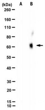
Western Blotting Analysis: A representative lot detected minicore HCV in Western Blotting applications (Eng, F.J., et. al. (2018). Hepatol Commun. 2(1):21-28).
Immunofluorescence Analysis: A representative lot detected minicore HCV in Immunofluorescence applications (Eng, F.J., et. al. (2018). Hepatol Commun. 2(1):21-28).
Quality
Evaluated by Western Blotting in extracts of HEK293T cells transfected with plasmids expressing only HCV p21 core and 91 minicore proteins.
Western Blotting Analysis: 4 ug/mL of this antibody detected minicore HCV in extracts of HEK293T cells transfected with plasmids expressing only HCV p21 core and 91 minicore proteins.
Target description
~ 8 and 21 kDa observed. Uncharacterized bands may be observed in some lysate(s).
Physical form
Format: Purified
Protein G purified
Purified mouse monoclonal antibody IgG1 in buffer containing 0.1 M Tris-Glycine (pH 7.4), 150 mM NaCl with 0.05% sodium azide.
Storage and Stability
Stable for 1 year at 2-8°C from date of receipt.
Other Notes
Concentration: Please refer to lot specific datasheet.
Disclaimer
Unless otherwise stated in our catalog or other company documentation accompanying the product(s), our products are intended for research use only and are not to be used for any other purpose, which includes but is not limited to, unauthorized commercial uses, in vitro diagnostic uses, ex vivo or in vivo therapeutic uses or any type of consumption or application to humans or animals.


