Skip to Content
Merck
All Photos(4)

Key Documents

HPA015270

Sigma-Aldrich

Anti-STK4 antibody produced in rabbit

enhanced validation

Prestige Antibodies® Powered by Atlas Antibodies, affinity isolated antibody, buffered aqueous glycerol solution

Synonym(s):

Anti-MST-1, Anti-Mammalian STE20-like protein kinase 1, Anti-STE20-like kinase MST1, Anti-Serine/threonine-protein kinase 4, Anti-Serine/threonine-protein kinase Krs-2

Sign Into View Organizational & Contract Pricing


About This Item

UNSPSC Code:
12352203
Human Protein Atlas Number:
NACRES:
NA.41

biological source

rabbit

Quality Level

conjugate

unconjugated

antibody form

affinity isolated antibody

antibody product type

primary antibodies

clone

polyclonal

product line

Prestige Antibodies® Powered by Atlas Antibodies

form

buffered aqueous glycerol solution

species reactivity

rat, mouse, human

enhanced validation

RNAi knockdown
Learn more about Antibody Enhanced Validation

technique(s)

immunoblotting: 0.04-0.4 μg/mL
immunofluorescence: 0.25-2 μg/mL
immunohistochemistry: 1:50-1:200

immunogen sequence

IEHDDTLPSQLGTMVINAEDEEEEGTMKRRDETMQPAKPSFLEYFEQKEKENQINSFGKSVPGPLKNSSDWKIPQDGDYEF

UniProt accession no.

shipped in

wet ice

storage temp.

−20°C

target post-translational modification

unmodified

Gene Information

human ... STK4(6789)

General description

STK4 (serine/threonine kinase 4) is a kinase protein, which is ubiquitous in nature. It is homologous to yeast Ste20 and Drosophila Hippo protein. It has a molecular weight of 63kDa, and is a resident of the cytoplasm. It is also called MST1 (mammalian sterile 20-like protein), and has its active site at its N-terminal. It also contains an autoinhibitory domain, and a coiled-coil SARAH domain at its C-terminal. This domain is responsible for protein hetero- and homodimerization.

Immunogen

Serine/threonine-protein kinase 4 recombinant protein epitope signature tag (PrEST)

Application

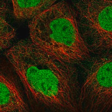
Anti-STK4 antibody produced in rabbit, a Prestige Antibody, is developed and validated by the Human Protein Atlas (HPA) project . Each antibody is tested by immunohistochemistry against hundreds of normal and disease tissues. These images can be viewed on the Human Protein Atlas (HPA) site by clicking on the Image Gallery link. The antibodies are also tested using immunofluorescence and western blotting. To view these protocols and other useful information about Prestige Antibodies and the HPA, visit sigma.com/prestige.

Biochem/physiol Actions

STK4 (serine/threonine kinase 4) functions as both apoptotic and anti-apoptotic protein. It acts as an apoptotic protein upon its cleavage by caspases, which produces an N-terminal fragment of 36kDa. This fragment moves to the nucleus, where it phosphorylates histone proteins, resulting in apoptosis initiation. It also interacts with JNK (Jun N-terminal kinase) and RASSF1A (Ras association domain family member 1) to induce apoptosis. It phosphorylates FOXO1 (forkhead box O1) and FOXO3, which are transcription factors of FOXO family, in stress-response pathway. Thus, it maintains the viability of cell. Inactivation of this gene in lymphocytes and neutrophils leads to abnormal mitochondrial membrane potential. This results in increased chances of apoptotic death. Thus, loss of this gene, in humans is a cause of primary immunodeficiency syndrome. It is essential for the homing and maintenance of naïve T-cells, and deficiency in this gene leads to abnormal development and/or maintenance of regulatory T-cells.

Features and Benefits

Prestige Antibodies® are highly characterized and extensively validated antibodies with the added benefit of all available characterization data for each target being accessible via the Human Protein Atlas portal linked just below the product name at the top of this page. The uniqueness and low cross-reactivity of the Prestige Antibodies® to other proteins are due to a thorough selection of antigen regions, affinity purification, and stringent selection. Prestige antigen controls are available for every corresponding Prestige Antibody and can be found in the linkage section.

Every Prestige Antibody is tested in the following ways:
  • IHC tissue array of 44 normal human tissues and 20 of the most common cancer type tissues.
  • Protein array of 364 human recombinant protein fragments.

Linkage

Corresponding Antigen APREST73444

Physical form

Solution in phosphate-buffered saline, pH 7.2, containing 40% glycerol and 0.02% sodium azide

Legal Information

Prestige Antibodies is a registered trademark of Merck KGaA, Darmstadt, Germany

Disclaimer

Unless otherwise stated in our catalog or other company documentation accompanying the product(s), our products are intended for research use only and are not to be used for any other purpose, which includes but is not limited to, unauthorized commercial uses, in vitro diagnostic uses, ex vivo or in vivo therapeutic uses or any type of consumption or application to humans or animals.

Not finding the right product?  

Try our Product Selector Tool.

Storage Class Code

10 - Combustible liquids

WGK

WGK 1

Flash Point(F)

Not applicable

Flash Point(C)

Not applicable

Personal Protective Equipment

dust mask type N95 (US), Eyeshields, Gloves

Certificates of Analysis (COA)

Search for Certificates of Analysis (COA) by entering the products Lot/Batch Number. Lot and Batch Numbers can be found on a product’s label following the words ‘Lot’ or ‘Batch’.

Already Own This Product?

Find documentation for the products that you have recently purchased in the Document Library.

Visit the Document Library

Nadine T Nehme et al.
Blood, 119(15), 3458-3468 (2011-12-17)
The molecular mechanisms that underlie T-cell quiescence are poorly understood. In the present study, we report a primary immunodeficiency phenotype associated with MST1 deficiency and primarily characterized by a progressive loss of naive T cells. The in vivo consequences include
Ingrid Babel et al.
Molecular & cellular proteomics : MCP, 10(3), M110-M110 (2011-01-14)
The characterization of the humoral response in cancer patients is becoming a practical alternative to improve early detection. We prepared phage microarrays containing colorectal cancer cDNA libraries to identify phage-expressed peptides recognized by tumor-specific autoantibodies from patient sera. From a
Hengameh Abdollahpour et al.
Blood, 119(15), 3450-3457 (2012-02-02)
We describe a novel clinical phenotype associating T- and B-cell lymphopenia, intermittent neutropenia, and atrial septal defects in 3 members of a consanguineous kindred. Their clinical histories included recurrent bacterial infections, viral infections, mucocutaneous candidiasis, cutaneous warts, and skin abscesses.
Igor Govorov et al.
Scientific reports, 12(1), 22154-22154 (2022-12-23)
In a previous study, we showed that serine/threonine-protein kinase 4 (STK4) is involved in the control on proliferation and migration of endometrial cancer (EC) cells in vitro. In the present paper, we studied STK4 expression in EC tissues from a
Jiujie Cui et al.
Molecular cancer research : MCR, 17(6), 1316-1325 (2019-02-24)
Pancreatic ductal adenocarcinoma (PDAC) is a deadly disease, and its incidence is increasing annually. It is critical to reveal and delineate the molecular mechanism promoting PDAC development and progression. Mammalian STE20-like kinase 1 (MST1) is a proapoptotic cytoplasmic kinase and

Our team of scientists has experience in all areas of research including Life Science, Material Science, Chemical Synthesis, Chromatography, Analytical and many others.

Contact Technical Service