Skip to Content
Merck

C5992

Sigma-Aldrich

Anti-Cytokeratin, pan antibody, Mouse monoclonal

clone PCK-26, purified from hybridoma cell culture

Synonym(s):

Monoclonal Anti-pan-Cytokeratin, Anti-Cytokeratin, Anti-Cytokeratin - Monoclonal Anti-Cytokeratin, pan antibody produced in mouse

Sign Into View Organizational & Contract Pricing


About This Item

UNSPSC Code:
12352203
NACRES:
NA.41

Pricing and availability is not currently available.

biological source

mouse

Quality Level

conjugate

unconjugated

antibody form

purified from hybridoma cell culture

antibody product type

primary antibodies

clone

PCK-26, monoclonal

form

buffered aqueous solution

species reactivity

human, chicken, snake, hamster, pig, goat, feline, bovine, carp, rat, rabbit, canine, lizard, sheep, mouse, guinea pig

packaging

antibody small pack of 25 μL

concentration

~1.5 mg/mL

Looking for similar products? Visit Product Comparison Guide

General description

Monoclonal Anti-Pan Cytokeratin (mouse IgG1 isotype) is derived from the PCK-26 hybridoma produced by the fusion of mouse myeloma cells and splenocytes from BALB/c mice immunized with a cytokeratin preparation from human epidermis. Cytokeratins are a group of at least 29 different proteins. They are characteristic of epithelial and trichocytic cells. Cytokeratin peptide 1 (68 kDa) is expressed together with cytokeratin 10 in the suprabasal cell layers or the differentiation compartment of the epidermis. Its expression increases with epidermal maturation and is modified post translationally in terminally differentiated keratinocytes of the stratum corneum. Cytokeratin peptide 5 (58 kDa) is the primary type II keratin in stratified epithelia, while cytokeratin type 8 (52 kDa) is a major type II keratin in simple epithelia. Cytokeratin 6 (56 kDa) is a "hyperproliferation" cytokeratin expressed in tissues with natural or pathological high turnover. Monoclonal antibodies to cytokeratins are specific markers of epithelial cell differentiation and have been widely used as tools in tumor identification and classification.

Specificity

The antibody recognizes an epitope located on the Type II cytokeratins 1, 5, 6, and 8. PCK-26 is a broad spectrum antibody which reacts specifically with a variety of normal, reactive, and neoplastic epithelial tissues. The antibody reacts with simple, cornifying, and non-cornifying squamous epithelia and pseudostratified epithelia.

Immunogen

cytokeratin from human epidermis.

Application

Applications in which this antibody has been used successfully, and the associated peer-reviewed papers, are given below.
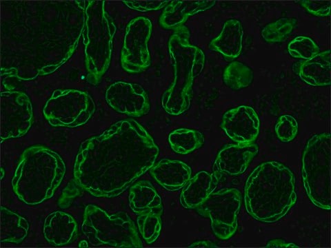
Immunofluorescence (1 paper)
Monoclonal Anti-Cytokeratin, pan antibody produced in mouse has been used in:
  • immunoblotting
  • immunohistochemistry
  • Immunocytochemistry
  • dot blotting

Biochem/physiol Actions

Cytokeratin facilitates the typing of normal, metaplastic, and neoplastic cells and may aid in the discrimination of carcinomas and non-epithelial tumors such as sarcomas, lymphomas, and neural tumors. It is also useful in detecting micrometastases in lymph nodes and other tissues, and for determining the origin of poorly differentiated tumors.

Physical form

Solution in 0.01 M phosphate buffered saline, pH 7.4, containing 15 mM sodium azide.

Disclaimer

Unless otherwise stated in our catalog or other company documentation accompanying the product(s), our products are intended for research use only and are not to be used for any other purpose, which includes but is not limited to, unauthorized commercial uses, in vitro diagnostic uses, ex vivo or in vivo therapeutic uses or any type of consumption or application to humans or animals.

Not finding the right product?  

Try our Product Selector Tool.

Storage Class Code

12 - Non Combustible Liquids

WGK

WGK 1

Flash Point(F)

Not applicable

Flash Point(C)

Not applicable


Certificates of Analysis (COA)

Search for Certificates of Analysis (COA) by entering the products Lot/Batch Number. Lot and Batch Numbers can be found on a product’s label following the words ‘Lot’ or ‘Batch’.

Need A Sample COA?

This is a sample Certificate of Analysis (COA) and may not represent a recently manufactured lot of this specific product.

Already Own This Product?

Find documentation for the products that you have recently purchased in the Document Library.

Visit the Document Library

Marta Campillo Poveda et al.
STAR protocols, 4(4), 102608-102608 (2023-09-26)
Parasitic helminth worms frequently infect the gastrointestinal tract and interact with the intestinal epithelium and specialized cell types within it. Intestinal organoids derived from stem cells that line the intestine represent a transformational technology in the study of epithelial-parasite dialogue.
Nasser K Altorki et al.
Cell reports. Medicine, 5(3), 101438-101438 (2024-02-25)
In early-stage non-small cell lung cancer, the combination of neoadjuvant anti-PD-L1 and subablative stereotactic body radiation therapy (SBRT) is associated with higher rates of major pathologic response compared to anti-PD-L1 alone. Here, we identify a 140-gene set, enriched in genes
Martin Kann et al.
Development (Cambridge, England), 142(7), 1254-1266 (2015-03-26)
Development of the metanephric kidney depends on tightly regulated interplay between self-renewal and differentiation of a nephron progenitor cell (NPC) pool. Several key factors required for the survival of NPCs have been identified, including fibroblast growth factor (FGF) signaling and
Nasser K Altorki et al.
Cell reports, 39(1), 110639-110639 (2022-04-07)
To investigate changes in the tumor microenvironment (TME) during lung cancer progression, we interrogate tumors from two chest computed tomography (CT)-defined groups. Pure non-solid (pNS) CT density nodules contain preinvasive/minimally invasive cancers, and solid density nodules contain invasive cancers. Profiling
Alison Min-Yan Cheung et al.
Breast cancer research : BCR, 23(1), 114-114 (2021-12-20)
The extent of cellular heterogeneity in breast cancer could have potential impact on diagnosis and long-term outcome. However, pathology evaluation is limited to biomarker immunohistochemical staining and morphology of the bulk cancer. Inter-cellular heterogeneity of biomarkers is not usually assessed.

Our team of scientists has experience in all areas of research including Life Science, Material Science, Chemical Synthesis, Chromatography, Analytical and many others.

Contact Technical Service