Skip to Content
Merck
All Photos(3)

Key Documents

C3237

Sigma-Aldrich

Anti-Caveolin-1 antibody produced in rabbit

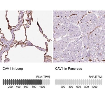
enhanced validation

IgG fraction of antiserum, buffered aqueous solution

Synonym(s):

Anti-Caveolin-1, Anti-VIP21

Sign Into View Organizational & Contract Pricing


About This Item

MDL number:
UNSPSC Code:
12352203
NACRES:
NA.41

biological source

rabbit

Quality Level

conjugate

unconjugated

antibody form

IgG fraction of antiserum

antibody product type

primary antibodies

clone

polyclonal

form

buffered aqueous solution

mol wt

antigen 22 kDa

species reactivity

human, rat, mouse

enhanced validation

independent
Learn more about Antibody Enhanced Validation

technique(s)

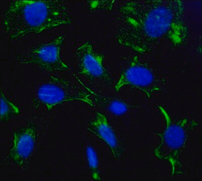
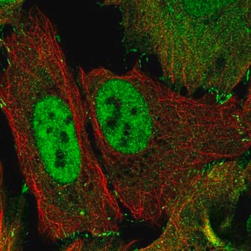
indirect immunofluorescence: 1:500 using mouse fibroblast NIH3T3 cell line
microarray: suitable
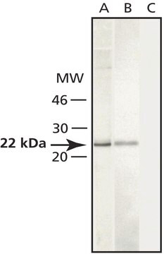
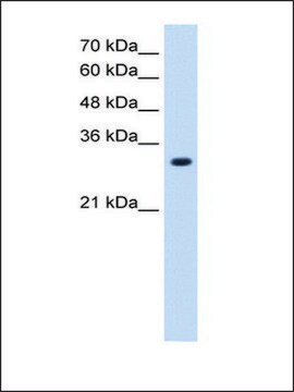
western blot: 1:2,000 using whole cell extract of the human umbilical vein endothelial HUVEC cell line and the rat fibroblast Rat-1 cell line

UniProt accession no.

shipped in

dry ice

storage temp.

−20°C

target post-translational modification

unmodified

Gene Information

human ... CAV1(857)
mouse ... Cav1(12389)
rat ... Cav1(25404)

Looking for similar products? Visit Product Comparison Guide

General description

Anti-Caveolin-1 is developed in rabbit using a synthetic peptide corresponding to a region at the N-terminus of human caveolin-1 conjugated to keyhole limpet hemocyanin (KLH) as immunogen. Caveolin-1 (CAV1) is a multifunctional scaffolding protein, that has three exons. It is located on chromosome 7q31.
Caveolin, a 20-24-kDa integral transmembrane protein, has been identified as a principal component of caveolae membranes. Caveolin (also termed VIP21) exists in several isoforms termed caveolin-1, caveolin-2 and caveolin-3. Caveolin-1 (20-22 kDa) can exist as two isoforms, caveolin-1α and -1β due to alternative splicing of the mRNA.

Specificity

The sequence is highly conserved in many species, e.g., identical in rat, mouse, bovine, and dog caveolin-1. The sequence is not found in caveolin-2 or caveolin-3.

Immunogen

synthetic peptide corresponding to a region at the N-terminus of human caveolin-1 (amino acids 2-20).

Application

Anti-Caveolin-1 antibody produced in rabbit has been used in:
  • indirect immunofluorescence
  • in confocal microscopy
  • western blotting

Biochem/physiol Actions

Caveolin family members function as scaffolding proteins to organize and concentrate specific lipids such as cholesterol and glycosylphosphatidylinositol (GPI) and lipid modified signaling molecules within caveolae membranes. Staining of caveolin-1 in immunoblotting is specifically inhibited with caveolin-1 immunizing peptide (human, amino acids 2-20).†
Caveolin-1 is expected to play a major role in the growth and evolution of cancer. It participates in glucose metabolism. It may also participate in angiogenesis. Cav-1 helps to control the expression of glucose transporters.

Physical form

Solution in 0.01 M phosphate buffered saline, pH 7.4, containing 15 mM sodium azide.

Disclaimer

Unless otherwise stated in our catalog or other company documentation accompanying the product(s), our products are intended for research use only and are not to be used for any other purpose, which includes but is not limited to, unauthorized commercial uses, in vitro diagnostic uses, ex vivo or in vivo therapeutic uses or any type of consumption or application to humans or animals.

Not finding the right product?  

Try our Product Selector Tool.

Storage Class Code

10 - Combustible liquids

WGK

WGK 1

Flash Point(F)

Not applicable

Flash Point(C)

Not applicable


Certificates of Analysis (COA)

Search for Certificates of Analysis (COA) by entering the products Lot/Batch Number. Lot and Batch Numbers can be found on a product’s label following the words ‘Lot’ or ‘Batch’.

Already Own This Product?

Find documentation for the products that you have recently purchased in the Document Library.

Visit the Document Library

Rac1 participates in thermally induced alterations of the cytoskeleton, cell morphology and lipid rafts, and regulates the expression of heat shock proteins in B16F10 melanoma cells
Gungor B, et al.
Testing, 9(2), e89136-e89136 (2014)
Co-expression of MG29 and Ryanodine Receptor Leads to Apoptotic Cell Death EFFECT MEDIATED BY INTRACELLULAR Ca2+ RELEASE
Pan Z, et al.
The Journal of Biological Chemistry, 279(19), 19387-19390 (2004)
Camila de Souza Palma et al.
Molecular & cellular proteomics : MCP, 15(3), 906-917 (2016-01-15)
Epithelial to mesenchymal transition (EMT)(1) occurs naturally during embryogenesis, tissue repair, cancer progression, and metastasis. EMT induces cellular and microenvironmental changes resulting in loss of epithelial and acquisition of mesenchymal phenotypes, which promotes cellular invasive and migratory capabilities. EMT can
Burcin Gungor et al.
PloS one, 9(2), e89136-e89136 (2014-03-04)
Eukaryotic cells exhibit a characteristic response to hyperthermic treatment, involving morphological and cytoskeletal alterations and the induction of heat shock protein synthesis. Small GTPases of the Ras superfamily are known to serve as molecular switches which mediate responses to extracellular
Jennifer van der Horst et al.
The Journal of general physiology, 153(3) (2021-02-04)
The dynein motor protein transports proteins away from the cell membrane along the microtubule network. Recently, we found the microtubule network was important for regulating the membrane abundance of voltage-gated Kv7.4 potassium channels in vascular smooth muscle. Here, we aimed

Our team of scientists has experience in all areas of research including Life Science, Material Science, Chemical Synthesis, Chromatography, Analytical and many others.

Contact Technical Service