Skip to Content
Merck
All Photos(2)

Key Documents

C8791

Sigma-Aldrich

Monoclonal Anti-Cytokeratin Peptide 14 antibody produced in mouse

clone CKB1, ascites fluid

Synonym(s):

Anti-CK14, Anti-EBS1, Anti-EBS1A, Anti-EBS1B, Anti-EBS1C, Anti-EBS1D, Anti-EBS3, Anti-EBS4, Anti-K14, Anti-NFJ

Sign Into View Organizational & Contract Pricing


About This Item

MDL number:
UNSPSC Code:
12352203
NACRES:
NA.41

biological source

mouse

Quality Level

conjugate

unconjugated

antibody form

ascites fluid

antibody product type

primary antibodies

clone

CKB1, monoclonal

contains

15 mM sodium azide

species reactivity

human

technique(s)

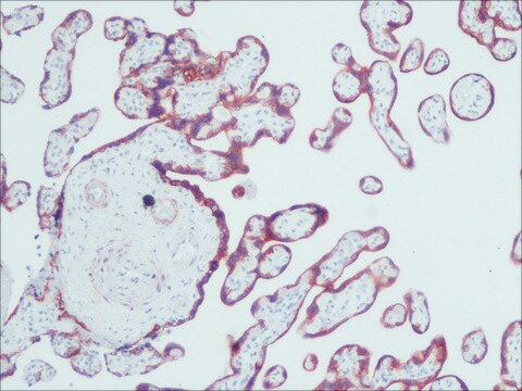
immunohistochemistry (formalin-fixed, paraffin-embedded sections): suitable
immunohistochemistry (frozen sections): suitable
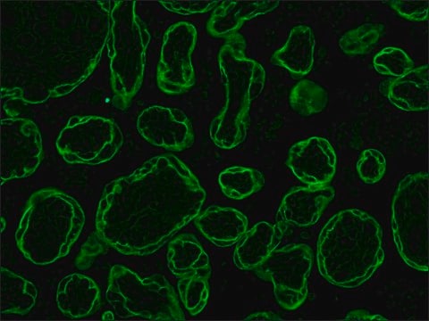
indirect immunofluorescence: 1:200 using methacarn-fixed, paraffin-embedded human tissue sections

isotype

IgM

UniProt accession no.

shipped in

dry ice

storage temp.

−20°C

target post-translational modification

unmodified

Gene Information

human ... KRT14(3861)

General description

Monoclonal Anti-Cytokeratin Peptide 14 (mouse IgM isotype) is derived from the hybridoma produced by the fusion of mouse myeloma cells and splenocytes from an immunized mouse. Keratin 14 (KRT14) is expressed in keratinocytes and localized to the basal epidermal layer. The gene is mapped to human chromosome 17q21 and encodes an intermediate filament.

Specificity

The antibody reacts with myoepithelial cells in various organs (parotid, submandibular, mammary, prostate, and sweat glands) as well as with the basal layer of certain squamous epithelium (exocervical, laryngeal epithelium).

Immunogen

human callus keratins.

Application

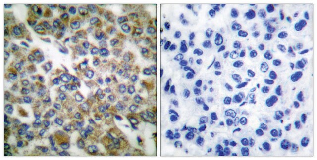
Monoclonal Anti-Cytokeratin Peptide 14 antibody produced in mouse has been used in indirect immunofluorescence or immunoperoxidase techniques.

Biochem/physiol Actions

Keratin 14 (KRT14) is associated with maintaining cellular stability. Mutation in the KRT14 gene leads to epidermolysis bullosa simplex, a genetic disorder. It is known to promote angiogenesis, cell migration and proliferation in cancer cells.

Disclaimer

Unless otherwise stated in our catalog or other company documentation accompanying the product(s), our products are intended for research use only and are not to be used for any other purpose, which includes but is not limited to, unauthorized commercial uses, in vitro diagnostic uses, ex vivo or in vivo therapeutic uses or any type of consumption or application to humans or animals.

Not finding the right product?  

Try our Product Selector Tool.

Storage Class Code

10 - Combustible liquids

WGK

nwg

Flash Point(F)

Not applicable

Flash Point(C)

Not applicable


Certificates of Analysis (COA)

Search for Certificates of Analysis (COA) by entering the products Lot/Batch Number. Lot and Batch Numbers can be found on a product’s label following the words ‘Lot’ or ‘Batch’.

Already Own This Product?

Find documentation for the products that you have recently purchased in the Document Library.

Visit the Document Library

Y Gache et al.
The Journal of clinical investigation, 97(10), 2289-2298 (1996-05-15)
Epidermolysis bullosa simplex with muscular dystrophy (MD-EBS) is a disease characterized by generalized blistering of the skin associated with muscular involvement. We report that the skin of three MD-EBS patients is not reactive with antibodies 6C6, 10F6, or 5B3 raised
Amina Errazahi et al.
Journal of bone and mineral research : the official journal of the American Society for Bone and Mineral Research, 18(4), 737-750 (2003-04-04)
The presence of identical or distinct type I parathyroid hormone (PTH)/parathyroid hormone-related peptide (PTHrP) receptors in keratinocytes is still a matter of debate. We studied the expression and functionality of PTHrP receptors in freshly isolated keratinocytes from newborn rat skin.
Cut and Paste: Efficient Homology-Directed Repair of a Dominant Negative KRT14 Mutation via CRISPR/Cas9 Nickases
Kocher, et al.
Molecular Therapy, 25(11), 2585-2585 (2017)
Maja Matic et al.
Methods in molecular biology (Clifton, N.J.), 289, 193-200 (2004-10-27)
Single-cell suspensions of primary keratinocytes comprise a heterogeneous cell population that consists of basal cells (stem cells, transient amplifying cells, and post-mitotic basal cells) and suprabasal cells at different stages of differentiation. Quantitative data for the differential expression of epitopes
Sally Dabelsteen et al.
Stem cells (Dayton, Ohio), 27(6), 1388-1399 (2009-06-03)
Human embryonic stem (hES) cells can generate cells expressing p63, K14, and involucrin, which have been proposed to be keratinocytes. Although these hES-derived, keratinocyte-like (hESderK) cells form epithelioid colonies when cultured in a fibroblast feeder system optimal for normal tissue-derived

Our team of scientists has experience in all areas of research including Life Science, Material Science, Chemical Synthesis, Chromatography, Analytical and many others.

Contact Technical Service