Skip to Content
Merck
All Photos(2)

Key Documents

G2404

Sigma-Aldrich

Anti-Golgi FTCD antibody, clone 58k-9, Mouse monoclonal

ascites fluid

Synonym(s):

Anti-LCHC1

Sign Into View Organizational & Contract Pricing


About This Item

MDL number:
UNSPSC Code:
12352203
NACRES:
NA.41

biological source

mouse

Quality Level

conjugate

unconjugated

antibody form

ascites fluid

antibody product type

primary antibodies

mol wt

antigen 58 kDa

contains

15 mM sodium azide

species reactivity

human, bovine, mouse, pig, canine, kangaroo rat, hamster, monkey, rat

technique(s)

electron microscopy: suitable
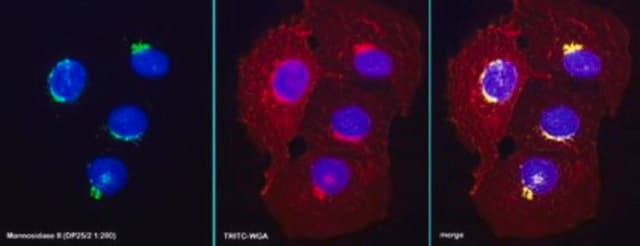
indirect immunofluorescence: 1:50 using cultured CHO cells
western blot: 1:5,000 using whole rat liver extract

isotype

IgG1

UniProt accession no.

shipped in

dry ice

storage temp.

−20°C

target post-translational modification

unmodified

Gene Information

human ... FTCD(10841)
mouse ... Ftcd(14317)
rat ... Ftcd(89833)

General description

FTCD is a homooctamer with its subunits arranged in a planar ring. It is predominantly expressed in the liver. Apart from being associated with the Golgi apparatus, it is detectable in the supernatant cytosolic fraction, and may also be localized to cytoplasmic vesicles.
Monoclonal Anti-Golgi 58K Protein/formiminotransferase cyclodeaminase (FTCD) (mouse IgG1 isotype) is derived from the 58K-9 hybridoma produced by the fusion of mouse myeloma cells and splenocytes from a mouse immunized with FTCD/Golgi 58K protein purified from rat liver.

Specificity

The antibody recognizes an epitope located on the microtubule-binding peripheral Golgi membrane 58 kDa protein. It is also useful for studies on the effect of microtubule-perturbing agents on the Golgi apparatus.

Immunogen

Golgi 58K protein from rat liver.

Application

Anti-Golgi FTCD antibody, clone 58k-9, Mouse monoclonal has been used in western blot analysis and dual immunofluorescence staining.
Anti-Golgi FTCD antibody, clone 58k-9, Mouse monoclonal is suitable for electron microscopy, indirect immunofluorescence at a working dilution of 1:50 using cultured CHO cells and indirect immunoblotting at 1:5000 working dilution using whole rat liver extract. It is also useful for the localization of Golgi 58K protein using immunoblotting, dot blot, electron microscopy, and immunocytochemistry.
The antibody was used:


  • for the analysis of distribution of the 58K9 protein exclusively localized in the Golgi
  • as a primary antibody in the immunofluorescence analysis in studies related to functioning of nuclear envelope protein TMEM209 in lung carcinoma cells, tracking TG2 (Transglutaminase Type 2) transport in renal tubular epithelial cells, binding of TRADD (TNFR-associated death domain protein) to TNF-R1 at the plasma membrane and localization of Wilson disease protein in the Golgi apparatus
  • in immunoprecipitation studies
Applications in which this antibody has been used successfully, and the associated peer-reviewed papers, are given below.
Immunofluorescence (1 paper)
Western Blotting (1 paper)

Biochem/physiol Actions

FTCD/58K protein of the Golgi apparatus used in conjunction with other antibodies to Golgi proteins (e.g., the Golgi β-COP protein) may be used for studies on the role and relationships of this protein in the cell. It is also useful for localization studies following subcellular fractionation procedures.
Golgi 58K protein is associated with the Golgi apparatus peripherally and has been identified as a version of FTCD (Formiminotransferase Cyclodeaminase). It is a bifunctional metabolic enzyme involved in conversion of histidine to glutamic acid. It acts as a channel for the transport of one-carbon units from formiminoglutamate to the folate pool. It also interacts with vimentin subunits and with polymerized vimentin filaments. Autosomal recessive disorder glutamate formiminotransferase deficiency is caused by defects in FTCD.

Disclaimer

Unless otherwise stated in our catalog or other company documentation accompanying the product(s), our products are intended for research use only and are not to be used for any other purpose, which includes but is not limited to, unauthorized commercial uses, in vitro diagnostic uses, ex vivo or in vivo therapeutic uses or any type of consumption or application to humans or animals.

Not finding the right product?  

Try our Product Selector Tool.

Storage Class Code

12 - Non Combustible Liquids

WGK

nwg

Flash Point(F)

Not applicable

Flash Point(C)

Not applicable


Certificates of Analysis (COA)

Search for Certificates of Analysis (COA) by entering the products Lot/Batch Number. Lot and Batch Numbers can be found on a product’s label following the words ‘Lot’ or ‘Batch’.

Already Own This Product?

Find documentation for the products that you have recently purchased in the Document Library.

Visit the Document Library

Human Cathepsin W, a Cysteine Protease Predominantly Expressed in NK Cells, Is Mainly Localized in the Endoplasmic Reticulum
Wex T, et al.
Journal of Immunology, 167(4), 2172-2178 (2001)
S J Jones et al.
Journal of immunology (Baltimore, Md. : 1950), 162(2), 1042-1048 (1999-01-23)
The subcellular localization of TNF-R1 to the Golgi apparatus, initially observed in endothelial cells, has been confirmed using transfection of bovine aortic endothelial cells with a human TNF-R1 expression plasmid. The subcellular interactions of TNF-R1 and the TRADD (TNFR-associated death
Ya-Sheng Gao et al.
European journal of cell biology, 81(7), 391-401 (2002-08-06)
Whether the highly dynamic structure of the vimentin intermediate filament (IF) cytoskeleton responds to cues from cellular organelles, and what proteins might participate in such events is largely unknown. We have shown previously that the Golgi protein formiminotransferase cyclodeaminase (FTCD)
Jonathan H Clarke et al.
The Journal of comparative neurology, 517(3), 296-312 (2009-09-17)
The role of cellular phosphatidylinositol 5-phosphate (PtdIns5P), as a signalling molecule or as a substrate for the production of small, compartmentalized pools of phosphatidylinositol 4,5-bisphosphate [PtdIns(4,5)P(2)], may be dependent on cell type and subcellular localization. PtdIns5P levels are primarily regulated
X L Yang et al.
The Biochemical journal, 326 ( Pt 3), 897-902 (1997-10-23)
Copper is an essential trace element in prokaryotes and eukaryotes and is strictly regulated by biological mechanisms. Menkes and Wilson diseases are human disorders that arise from disruption of the normal process of copper export from the cytosol to the

Our team of scientists has experience in all areas of research including Life Science, Material Science, Chemical Synthesis, Chromatography, Analytical and many others.

Contact Technical Service