Multiple epidermal growth factor-like domains protein 10 (Megf10) is a type 1 transmembrane protein with multiple EGF-like extracellular domains. Megf10 is highly expressed in the brain, and plays an important role in phacocytosis of apoptotic cells. This role may be performed through the interaction of Megf10 with the ATP-binding cassette transporter (ABCA1) protein. Previous studies have suggested that Megf10 may be involved in the uptake of amyloid β peptides in the brain. Megf10 may therefore play a role Alzheimer’s disease.
Specificity
Other homologies: Rat (98% sequence homology) and Human (92% sequence homology).
This antibody recognizes Megf10 at the cytoplasmic domain.
Immunogen
Epitope: Cytoplasmic domain
Histidine-tagged recombinant protein corresponding to the cytoplasmic domain of mouse Megf10.
Application
Detect the Megf10 protein using this Anti-Megf10 validated for use in WB & IF.
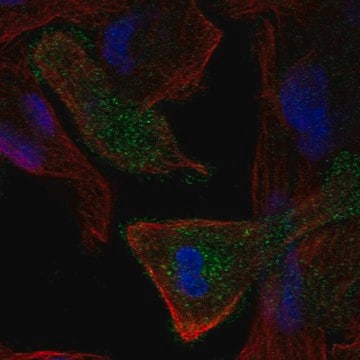
Immunofluorescence Analysis: A representative lot was used by an independent laboratory in P7 caudal brain tissue and retina cells. Image courtesy from the laboratory of Dr. Joshua Sanes, Harvard University.
Research Category Apoptosis & Cancer
Research Sub Category Apoptosis - Additional
Quality
Evaluated by Western Blot in mouse retina tissue lysate.
Western Blot Analysis: 1 µg/mL of this antibody detected Megf10 on 10 µg of mouse retina tissue lysate.
Target description
~130 kDa observed
Physical form
Affinity purified
Purified rabbit polyclonal in buffer containing 0.1 M Tris-Glycine (pH 7.4), 150 mM NaCl with 0.05% sodium azide.
Storage and Stability
Stable for 1 year at 2-8°C from date of receipt.
Analysis Note
Control Mouse retina tissue lysate
Other Notes
Concentration: Please refer to the Certificate of Analysis for the lot-specific concentration.
Disclaimer
Unless otherwise stated in our catalog or other company documentation accompanying the product(s), our products are intended for research use only and are not to be used for any other purpose, which includes but is not limited to, unauthorized commercial uses, in vitro diagnostic uses, ex vivo or in vivo therapeutic uses or any type of consumption or application to humans or animals.
Not finding the right product?
Try our Product Selector Tool.
Storage Class Code
12 - Non Combustible Liquids
WGK
WGK 1
Flash Point(F)
Not applicable
Flash Point(C)
Not applicable
Certificates of Analysis (COA)
Search for Certificates of Analysis (COA) by entering the products Lot/Batch Number. Lot and Batch Numbers can be found on a product’s label following the words ‘Lot’ or ‘Batch’.
Already Own This Product?
Find documentation for the products that you have recently purchased in the Document Library.
International journal of molecular sciences, 24(2) (2023-01-22)
It has been hypothesised that inhalational anaesthetics such as isoflurane (Iso) may trigger the pathogenesis of Alzheimer's disease (AD), while the gaseous anaesthetic xenon (Xe) exhibits many features of a putative neuroprotective agent. Loss of synapses is regarded as one
Microglia sculpt developing neural circuits by eliminating excess synapses in a process called synaptic pruning, by removing apoptotic neurons, and by promoting neuronal survival. To elucidate the role of microglia during embryonic and postnatal brain development, we used a mouse
The EMBO journal, 39(22), e104464-e104464 (2020-09-23)
Microglia are the principal phagocytes that clear cell debris in the central nervous system (CNS). This raises the question, which cells remove cell debris when microglial phagocytic activity is impaired. We addressed this question using Siglechdtr mice, which enable highly
International journal of molecular sciences, 25(2) (2024-01-23)
Synapse loss is one of the most critical features in Alzheimer's disease (AD) and correlates with cognitive decline. Astrocytes mediate synapse elimination through multiple EGF-like domains 10 (MEGF10) pathways in the developing and adult brain to build the precise neural
Frontiers in molecular neuroscience, 9, 105-105 (2016-11-09)
Cytoplasmic polyadenylation element binding protein 3 (CPEB3) regulates target RNA translation in neurons. Here, we examined CPEB3 distribution and function in the mouse retina. CPEB3 is expressed in retinal neurons, including those located in the inner nuclear layer (INL) and
Our team of scientists has experience in all areas of research including Life Science, Material Science, Chemical Synthesis, Chromatography, Analytical and many others.