Skip to Content
MilliporeSigma
All Photos(7)

Key Documents

HPA011294

Sigma-Aldrich

Anti-C1GALT1 antibody produced in rabbit

enhanced validation

Prestige Antibodies® Powered by Atlas Antibodies, affinity isolated antibody, buffered aqueous glycerol solution

Synonym(s):

Anti-Beta 1,3-galact, Anti-C1GalT1, Anti-Core 1 beta1,3-galactosyltransferase 1, Anti-Core 1 beta3-Gal-T, Anti-Core1 UDP- galactose:N-acetylgalactosamine-alpha-R beta 1,3-galactosyltransferase 1, Anti-Glycoprotein-N-acetylgalactosamine 3-beta-galactosyltransferase 1

Sign Into View Organizational & Contract Pricing

Select a Size

100 μL
$598.00

$598.00


Usually ships in 1 week. (Orders outside of US and Europe, please allow an additional 1-2 weeks for delivery)


Select a Size

Change View
100 μL
$598.00

About This Item

UNSPSC Code:
12352203
Human Protein Atlas Number:
NACRES:
NA.41

$598.00


Usually ships in 1 week. (Orders outside of US and Europe, please allow an additional 1-2 weeks for delivery)

biological source

rabbit

conjugate

unconjugated

antibody form

affinity isolated antibody

antibody product type

primary antibodies

clone

polyclonal

product line

Prestige Antibodies® Powered by Atlas Antibodies

form

buffered aqueous glycerol solution

species reactivity

human

enhanced validation

independent
Learn more about Antibody Enhanced Validation

technique(s)

immunohistochemistry: 1:20-1:50

immunogen sequence

VDTQPNVLHNDPHARHSDDNGQNHLEGQMNFNADSSQHKDENTDIAENLYQKVRILCWVMTGPQNLEKKAKHVKATWAQRCNKVLFMSSEENKDFPAVGLKTKEGRDQLYWKTIK

UniProt accession no.

shipped in

wet ice

storage temp.

−20°C

target post-translational modification

unmodified

Gene Information

human ... C1GALT1(56913)

General description

C1GALT1 (core 1 synthase, glycoprotein-N-acetylgalactosamine 3-β-galactosyltransferase 1) is a mucin-type O-glycosyltransferase, which resides in Golgi bodies. It is a type II transmembrane protein with its N-terminal facing the cytosol. Its transmembrane domain is made of 26 amino acids, and its functional domain is made of 331 amino acids and faces the lumen.

Immunogen

core 1 synthase, glycoprotein-N-acetylgalactosamine 3-beta-galactosyltransferase 1

Application

All Prestige Antibodies Powered by Atlas Antibodies are developed and validated by the Human Protein Atlas (HPA) project and as a result, are supported by the most extensive characterization in the industry.

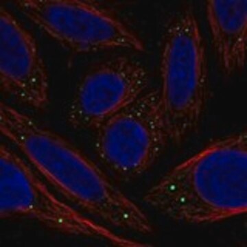
The Human Protein Atlas project can be subdivided into three efforts: Human Tissue Atlas, Cancer Atlas, and Human Cell Atlas. The antibodies that have been generated in support of the Tissue and Cancer Atlas projects have been tested by immunohistochemistry against hundreds of normal and disease tissues and through the recent efforts of the Human Cell Atlas project, many have been characterized by immunofluorescence to map the human proteome not only at the tissue level but now at the subcellular level. These images and the collection of this vast data set can be viewed on the Human Protein Atlas (HPA) site by clicking on the Image Gallery link. We also provide Prestige Antibodies® protocols and other useful information.

Biochem/physiol Actions

C1GALT1 (core 1 synthase, glycoprotein-N-acetylgalactosamine 3-β-galactosyltransferase 1) catalyzes the transfer of galactose (Gal) to N-acetylgalactosamine (GalNAc) of a serine (Ser) or threonine (Thr) residue (Tn antigen) to form the Galβ1-3GalNAcα1-Ser/Thr structure. This structure is called the T antigen or core 1 structure, which acts as the precursor for maturation of mucin-type O-glycans. This protein is also involved in the control of kidney development, thrombopoiesis and angiogenesis. It is up-regulated in hepatocellular carcinoma (HCC) and is responsible for enhanced proliferation of HCC cells. This is achieved by the activation of hepatocyte growth factor (HGF) by C1GALT1, through modulation of MET O-glycosylation. It is responsible for the enhanced invasiveness of colon cancer via the modification of the O-glycosylation of FGFR2 (fibroblast growth factor receptor 2).[1] Polymorphisms in C1GLAT1 gene are linked with Henoch-Schönlein purpura in Chinese population.

Features and Benefits

Prestige Antibodies® are highly characterized and extensively validated antibodies with the added benefit of all available characterization data for each target being accessible via the Human Protein Atlas portal linked just below the product name at the top of this page. The uniqueness and low cross-reactivity of the Prestige Antibodies® to other proteins are due to a thorough selection of antigen regions, affinity purification, and stringent selection. Prestige antigen controls are available for every corresponding Prestige Antibody and can be found in the linkage section.

Every Prestige Antibody is tested in the following ways:
  • IHC tissue array of 44 normal human tissues and 20 of the most common cancer type tissues.
  • Protein array of 364 human recombinant protein fragments.

Linkage

Corresponding Antigen APREST71747

Physical form

Solution in phosphate-buffered saline, pH 7.2, containing 40% glycerol and 0.02% sodium azide

Legal Information

Prestige Antibodies is a registered trademark of Merck KGaA, Darmstadt, Germany

Disclaimer

Unless otherwise stated in our catalog or other company documentation accompanying the product(s), our products are intended for research use only and are not to be used for any other purpose, which includes but is not limited to, unauthorized commercial uses, in vitro diagnostic uses, ex vivo or in vivo therapeutic uses or any type of consumption or application to humans or animals.

Not finding the right product?  

Try our Product Selector Tool.

Storage Class

10 - Combustible liquids

wgk_germany

WGK 1

flash_point_f

Not applicable

flash_point_c

Not applicable

ppe

Eyeshields, Gloves, multi-purpose combination respirator cartridge (US)


Choose from one of the most recent versions:

Certificates of Analysis (COA)

Lot/Batch Number

Don't see the Right Version?

If you require a particular version, you can look up a specific certificate by the Lot or Batch number.

Already Own This Product?

Find documentation for the products that you have recently purchased in the Document Library.

Visit the Document Library

JinDan An et al.
Rheumatology international, 33(10), 2539-2542 (2013-04-30)
Henoch-Schönlein purpura (HSP) is the most common systemic vasculitis of childhood. The molecular etiology of HSP is not well understood. The purpose of this study is to investigate the association between polymorphisms in C1GALT1 gene and the risk of HSP
Rajindra P Aryal et al.
The Journal of biological chemistry, 287(19), 15317-15329 (2012-03-15)
The interaction of the endoplasmic reticulum molecular chaperone Cosmc with its specific client T-synthase (Core 1 β1-3-galactosyltransferase) is required for folding of the enzyme and eventual movement of the T-synthase to the Golgi, but the mechanism of interaction is unclear.
Ji-Shiang Hung et al.
Oncotarget, 5(8), 2096-2106 (2014-04-25)
Core 1 β1,3-galactosyltransferase (C1GALT1) transfers galactose (Gal) to N-acetylgalactosamine (GalNAc) to form Galβ1,3GalNAc (T antigen). Aberrant O-glycans, such as T antigen, are commonly found in colorectal cancer. However, the role of C1GALT1 in colorectal cancer remains unclear. Here we showed
Yao-Ming Wu et al.
Cancer research, 73(17), 5580-5590 (2013-07-09)
Altered glycosylation is a hallmark of cancer. The core 1 β1,3-galactosyltransferase (C1GALT1) controls the formation of mucin-type O-glycans, far overlooked and underestimated in cancer. Here, we report that C1GALT1 mRNA and protein are frequently overexpressed in hepatocellular carcinoma tumors compared

Questions

Reviews

No rating value

Active Filters

Our team of scientists has experience in all areas of research including Life Science, Material Science, Chemical Synthesis, Chromatography, Analytical and many others.

Contact Technical Service