
HPA007338
Anti-RBP1 antibody produced in rabbit
affinity isolated antibody, buffered aqueous glycerol solution
Synonym(s):
Anti-CRABP-I, Anti-CRBP, Anti-CRBP1, Anti-CRBPI, Anti-RBPC
Select a Size
$850.00
$850.00
About This Item
Skip To
Product Name
Anti-RBP1 antibody produced in rabbit, affinity isolated antibody, buffered aqueous glycerol solution
biological source
rabbit
conjugate
unconjugated
antibody form
affinity isolated antibody
antibody product type
primary antibodies
clone
polyclonal
form
buffered aqueous glycerol solution
species reactivity
human
technique(s)
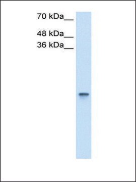
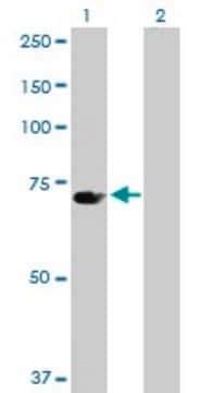
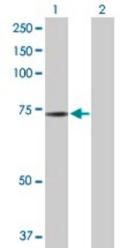
immunoblotting: 0.04-0.4 μg/mL
immunofluorescence: 0.25-2 μg/mL
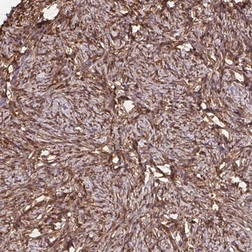
immunohistochemistry: 1:500-1:1000
immunogen sequence
MLVNENFEEYLRALDVNVALRKIANLLKPDKEIVQDGDHMIIRTLSTFRNYIMDFQVGKEFEEDLTGIDDRKCMTTVSWDGDKLQCVQKGEKEGRGWTQWIEGDELHLEMRVEGVVCKQVFKKVQ
UniProt accession no.
shipped in
wet ice
storage temp.
−20°C
target post-translational modification
unmodified
Quality Level
Gene Information
human ... RBP1(5947)
1 of 4
This Item | AV42081 | SAB1411379 | SAB1408678 |
|---|---|---|---|
| Quality Level 100 | Quality Level 100 | Quality Level 100 | Quality Level 100 |
| antibody form affinity isolated antibody | antibody form IgG fraction of antiserum | antibody form purified immunoglobulin | antibody form purified immunoglobulin |
| biological source rabbit | biological source rabbit | biological source rabbit | biological source rabbit |
| conjugate unconjugated | conjugate unconjugated | conjugate unconjugated | conjugate unconjugated |
| UniProt accession no. | UniProt accession no. | UniProt accession no. | UniProt accession no. |
| species reactivity human | species reactivity human, rabbit, guinea pig, rat, mouse, bovine, dog, horse | species reactivity human | species reactivity human |
Application
The Human Protein Atlas project can be subdivided into three efforts: Human Tissue Atlas, Cancer Atlas, and Human Cell Atlas. The antibodies that have been generated in support of the Tissue and Cancer Atlas projects have been tested by immunohistochemistry against hundreds of normal and disease tissues and through the recent efforts of the Human Cell Atlas project, many have been characterized by immunofluorescence to map the human proteome not only at the tissue level but now at the subcellular level. These images and the collection of this vast data set can be viewed on the Human Protein Atlas (HPA) site by clicking on the Image Gallery link. We also provide Prestige Antibodies® protocols and other useful information.
Disclaimer
Features and Benefits
Every Prestige Antibody is tested in the following ways:
- IHC tissue array of 44 normal human tissues and 20 of the most common cancer type tissues.
- Protein array of 364 human recombinant protein fragments.
Immunogen
Other Notes
Physical form
Legal Information
Not finding the right product?
Try our Product Selector Tool.
Storage Class
10 - Combustible liquids
wgk
WGK 1
flash_point_f
Not applicable
flash_point_c
Not applicable
Choose from one of the most recent versions:
Already Own This Product?
Find documentation for the products that you have recently purchased in the Document Library.
Active Filters
Our team of scientists has experience in all areas of research including Life Science, Material Science, Chemical Synthesis, Chromatography, Analytical and many others.
Contact Technical Service