Skip to Content
Merck
All Photos(1)

Documents

05-298

Sigma-Aldrich

Anti-α-Dystroglycan Antibody, clone VIA4-1

culture supernatant, clone VIA4-1, Upstate®

Synonym(s):

Anti-156DAG, Anti-A3a, Anti-AGRNR, Anti-DAG, Anti-LGMDR16, Anti-MDDGA9, Anti-MDDGC7, Anti-MDDGC9

Sign Into View Organizational & Contract Pricing


About This Item

UNSPSC Code:
12352203
eCl@ss:
32160702
NACRES:
NA.41

biological source

mouse

Quality Level

antibody form

culture supernatant

antibody product type

primary antibodies

clone

VIA4-1, monoclonal

species reactivity

guinea pig, rabbit, rat, human, mouse, canine

manufacturer/tradename

Upstate®

technique(s)

immunohistochemistry: suitable
western blot: suitable

isotype

IgG1

NCBI accession no.

UniProt accession no.

shipped in

wet ice

target post-translational modification

unmodified

Gene Information

human ... DAG1(1605)

Related Categories

General description

Dystroglycan is one of the dystrophin-associated glycoproteins, which is encoded by a 5.5 kb transcript in Homo sapiens by chromosome 3. There are two exons that are separated by a large intron. The spliced exons codes for a protein product is finally cleaved into two non-covalently associated subunits, α (N-terminal) and β (C-terminal). In skeletal muscle the dystroglycan complex works as a transmembrane linkage between the extracellular matrix and the cytoskeleton. α-dystroglycan is extracellular and binds to merosin (α-2 laminin) in the basement membrane, while β-dystroglycan is a transmembrane protein and binds to dystrophin, which is a large rod-like cytoskeletal protein, absent in Duchenne muscular dystrophy patients. Dystrophin binds to intracellular actin cables. In this way, the dystroglycan complex, which links the extracellular matrix to the intracellular actin cables, is thought to provide structural integrity in muscle tissues. The dystroglycan complex is also known to serve as an agrin receptor in muscle, where it may regulate agrin-induced acetylcholine receptor clustering at the neuromuscular junction. There is also evidence that suggests the function of dystroglycan as a part of the signal transduction pathway because it is shown that Grb2, a mediator of the Ras-related signal pathway, can interact with the cytoplasmic domain of dystroglycan. In general, aberrant expression of dystrophin-associated protein complex underlies the pathogenesis of Duchenne muscular dystrophy, Becker muscular dystrophy and severe childhood autosomal recessive muscular dystrophy. Interestingly, no genetic disease has been described for either α- or β-dystroglycan.
Dystroglycan is widely distributed in non-muscle tissues as well as in muscle tissues. During epithelial morphogenesis of kidney, the dystroglycan complex is shown to act as a receptor for the basement membrane. Dystroglycan expression in Mus musculus brain and neural retina has also been reported. However, the physiological role of dystroglycan in non-muscle tissues has remained unclear.
Note: This product may be used for research purposes only. Diagnostic use of this product requires a license from the University of Iowa Research Foundation, 214 Technology Innovation Center, Iowa City, IA 52242.

Specificity

Recognizes α-dystroglycan/LARGE-glycan.

Immunogen

Rabbit skeletal muscle membrane preparation

Application

Detect α-Dystroglycan using this Anti-α-Dystroglycan Antibody, clone VIA4-1. This antibody has been published and validated for use in IH and WB.
Research Category
Cell Structure
Research Sub Category
Cytoskeleton

Quality

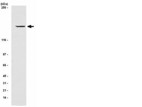
routinely evaluated on rabbit skeletal muscle in a western blot.

Target description

156 kDa

Physical form

Culture supernatant containing 0.05% sodium azide.
Unpurified

Storage and Stability

Maintain for 2 years at -20°C from date of shipment. Aliquot to avoid repeated freezing and thawing. For maximum recovery of product, centrifuge the original vial after thawing and prior to removing the cap.

Analysis Note

Control
Mouse, rat and human skeletal muscle tissue extracts or DU 145 (human prostate carcinoma tumor) cell lysate

Other Notes

Concentration: Please refer to the Certificate of Analysis for the lot-specific concentration.

Legal Information

UPSTATE is a registered trademark of Merck KGaA, Darmstadt, Germany

Disclaimer

Unless otherwise stated in our catalog or other company documentation accompanying the product(s), our products are intended for research use only and are not to be used for any other purpose, which includes but is not limited to, unauthorized commercial uses, in vitro diagnostic uses, ex vivo or in vivo therapeutic uses or any type of consumption or application to humans or animals.

Not finding the right product?  

Try our Product Selector Tool.

Storage Class Code

12 - Non Combustible Liquids

WGK

WGK 2


Certificates of Analysis (COA)

Search for Certificates of Analysis (COA) by entering the products Lot/Batch Number. Lot and Batch Numbers can be found on a product’s label following the words ‘Lot’ or ‘Batch’.

Already Own This Product?

Find documentation for the products that you have recently purchased in the Document Library.

Visit the Document Library

Dystrophin constitutes 5% of membrane cytoskeleton in skeletal muscle.
Ohlendieck, K and Campbell, K P
Febs Letters, 283, 230-234 (1991)
B J Collins et al.
Endocrinology, 142(7), 3152-3162 (2001-06-21)
Dystroglycan is a high affinity laminin-binding glycoprotein originally described as a member of the dystrophin-associated glycoprotein complex in muscle. We have demonstrated the presence of dystroglycan in the thyroid using immunocytochemistry, immunoblots, ligand binding assays, and relative quantitative RT-PCR. In
S Sugita et al.
The Journal of cell biology, 154(2), 435-445 (2001-07-27)
In nonneuronal cells, the cell surface protein dystroglycan links the intracellular cytoskeleton (via dystrophin or utrophin) to the extracellular matrix (via laminin, agrin, or perlecan). Impairment of this linkage is instrumental in the pathogenesis of muscular dystrophies. In brain, dystroglycan
Lara K Lee et al.
BMC neuroscience, 3, 10-10 (2002-08-17)
One of the earliest steps in synaptogenesis at the neuromuscular junction is the aggregation of nicotinic acetylcholine receptors at the postsynaptic membrane. This study presents quantitative analyses of receptor and alpha-Dystroglycan aggregation in response to agrin and laminin-1, alone or
Quiescence and activation of stem and precursor cell populations in the subependymal zone of the mammalian brain are associated with distinct cellular and extracellular matrix signals.
Kazanis, Ilias, et al.
The Journal of Neuroscience, 30, 9771-9781 (2010)

Our team of scientists has experience in all areas of research including Life Science, Material Science, Chemical Synthesis, Chromatography, Analytical and many others.

Contact Technical Service