Skip to Content
Merck
All Photos(1)

Key Documents

HPA001239

Sigma-Aldrich

Anti-MYH7 antibody produced in rabbit

Prestige Antibodies® Powered by Atlas Antibodies, affinity isolated antibody, buffered aqueous glycerol solution

Synonym(s):

MYH7 Antibody - Anti-MYH7 antibody produced in rabbit, Myh7 Antibody, Anti-MyHC-β antibody produced in rabbit, Anti-MyHC-slow antibody produced in rabbit, Anti-Myosin heavy chain 7 antibody produced in rabbit, Anti-Myosin heavy chain slow isoform antibody produced in rabbit, Anti-Myosin heavy chain, cardiac muscle β-isoform antibody produced in rabbit, Anti-Myosin-7 antibody produced in rabbit

Sign Into View Organizational & Contract Pricing


About This Item

UNSPSC Code:
12352203
Human Protein Atlas Number:
NACRES:
NA.41

biological source

rabbit

conjugate

unconjugated

antibody form

affinity isolated antibody

antibody product type

primary antibodies

clone

polyclonal

product line

Prestige Antibodies® Powered by Atlas Antibodies

form

buffered aqueous glycerol solution

species reactivity

human

technique(s)

immunohistochemistry: 1:1000- 1:2500

immunogen sequence

ALRKKHADSVAELGEQIDNLQRVKQKLEKEKSEFKLELDDVTSNMEQIIKAKANLEKMCRTLEDQMNEHRSKAEETQRSVNDLTSQRAKLQTENGELSRQLDEKEALISQLTRGKLTYTQQLEDLKRQLEEEVKAKNALAHALQS

UniProt accession no.

shipped in

wet ice

storage temp.

−20°C

target post-translational modification

unmodified

Gene Information

human ... MYH7(4625)

General description

Myosin heavy chain 7 (MYH7) comprises the N-terminal head domain and C-terminal rod domain. It also harbors the light meromyosin domain and actin-binding domain. The MYH7 gene is mapped to human chromosome location 14q11.2.

Immunogen

Myosin-7 recombinant protein epitope signature tag (PrEST)

Application

Anti-MYH7 antibody produced in rabbit has been used in:
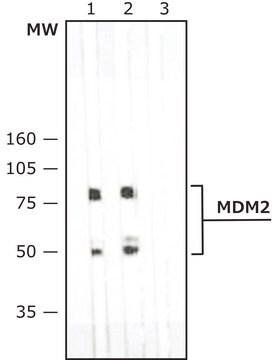
  • western blotting
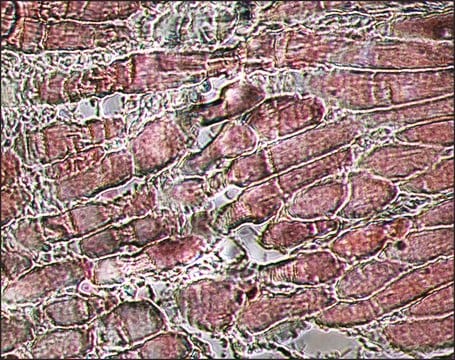
  • immunohistochemistry
  • immunofluorescence staining (1:100)

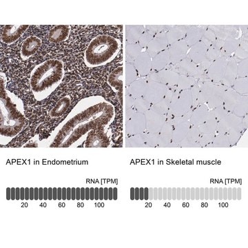
Anti-MYH7 antibody produced in rabbit is suitable for immunostaining to examine protein expression patterns in human cardiac sections using Protein Atlas confocal database.
Anti-MYH7 antibody produced in rabbit, a Prestige Antibody, is developed and validated by the Human Protein Atlas (HPA) project . Each antibody is tested by immunohistochemistry against hundreds of normal and disease tissues. These images can be viewed on the Human Protein Atlas (HPA) site by clicking on the Image Gallery link. The antibodies are also tested using immunofluorescence and western blotting. To view these protocols and other useful information about Prestige Antibodies and the HPA, visit sigma.com/prestige.

Biochem/physiol Actions

MYH7 (myosin, heavy chain 7, cardiac muscle, β) gene encodes the beta heavy chain subunit of cardiac myosin. It functions in muscle contraction. It is highly expressed in normal human ventricle and also in skeletal muscle tissues rich in slow-twitch type I muscle fibers. The expression of this protein is altered during thyroid hormone depletion and hemodynamic overloading. Defects in this gene cause familial hypertrophic cardiomyopathy, myosin storage myopathy, dilated cardiomyopathy, and Laing early-onset distal myopathy.

Features and Benefits

Prestige Antibodies® are highly characterized and extensively validated antibodies with the added benefit of all available characterization data for each target being accessible via the Human Protein Atlas portal linked just below the product name at the top of this page. The uniqueness and low cross-reactivity of the Prestige Antibodies® to other proteins are due to a thorough selection of antigen regions, affinity purification, and stringent selection. Prestige antigen controls are available for every corresponding Prestige Antibody and can be found in the linkage section.

Every Prestige Antibody is tested in the following ways:
  • IHC tissue array of 44 normal human tissues and 20 of the most common cancer type tissues.
  • Protein array of 364 human recombinant protein fragments.

Linkage

Corresponding Antigen APREST70519

Physical form

Solution in phosphate-buffered saline, pH 7.2, containing 40% glycerol and 0.02% sodium azide

Legal Information

Prestige Antibodies is a registered trademark of Merck KGaA, Darmstadt, Germany

Disclaimer

Unless otherwise stated in our catalog or other company documentation accompanying the product(s), our products are intended for research use only and are not to be used for any other purpose, which includes but is not limited to, unauthorized commercial uses, in vitro diagnostic uses, ex vivo or in vivo therapeutic uses or any type of consumption or application to humans or animals.

Not finding the right product?  

Try our Product Selector Tool.

Storage Class Code

10 - Combustible liquids

WGK

WGK 1

Flash Point(F)

Not applicable

Flash Point(C)

Not applicable

Personal Protective Equipment

dust mask type N95 (US), Eyeshields, Gloves

Certificates of Analysis (COA)

Search for Certificates of Analysis (COA) by entering the products Lot/Batch Number. Lot and Batch Numbers can be found on a product’s label following the words ‘Lot’ or ‘Batch’.

Already Own This Product?

Find documentation for the products that you have recently purchased in the Document Library.

Visit the Document Library

Hiroyuki Iwaki et al.
PloS one, 9(4), e88610-e88610 (2014-05-02)
MYH7 (also referred to as cardiac myosin heavy chain β) gene expression is known to be repressed by thyroid hormone (T3). However, the molecular mechanism by which T3 inhibits the transcription of its target genes (negative regulation) remains to be
Catherine E Alessi et al.
Journal of clinical neuromuscular disease, 22(1), 22-34 (2020-08-25)
Laing distal myopathy (LDM) is an autosomal dominant disorder caused by mutations in the slow skeletal muscle fiber myosin heavy chain (MYH7) gene on chromosome 14q11.2. The classic LDM phenotype-including early-onset, initial involvement of foot dorsiflexors and great toe extensors
Wenkun Dou et al.
Biosensors & bioelectronics, 175, 112875-112875 (2020-12-12)
The use of human induced pluripotent stem cell-derived cardiomyocytes (iPSC-CMs) as an in vitro model of the heart is limited by their structurally and functionally immature phenotypes. During heart development, mechanical stimuli from in vivo microenvironments are known to regulate
Chao Song et al.
Molecular therapy : the journal of the American Society of Gene Therapy, 29(3), 1120-1137 (2020-11-02)
Emerging evidence reveals that autophagy plays crucial roles in cardiac hypertrophy. Long noncoding RNAs (lncRNAs) are novel transcripts that function as gene regulators. However, it is unclear whether lncRNAs regulate autophagy in cardiac hypertrophy. Here, we identified a novel transcript
L Saez et al.
Nucleic acids research, 14(7), 2951-2969 (1986-04-11)
In an attempt to define myosin heavy chain (MHC) gene organization and expression in adult human skeletal muscle, we have isolated and characterized genomic sequences corresponding to different human sarcomeric MHC genes (1). In this report, we present the complete

Our team of scientists has experience in all areas of research including Life Science, Material Science, Chemical Synthesis, Chromatography, Analytical and many others.

Contact Technical Service