MABT889
Anti-CTHRC1 Antibody, clone Vli-55
clone Vli-55, from rabbit
Synonyme(s) :
Collagen triple helix repeat-containing protein 1, Protein NMTC1
About This Item
Produits recommandés
Source biologique
rabbit
Niveau de qualité
Forme d'anticorps
purified antibody
Type de produit anticorps
primary antibodies
Clone
Vli-55, monoclonal
Espèces réactives
rat, mouse, human, porcine
Technique(s)
immunohistochemistry: suitable
western blot: suitable
Numéro d'accès NCBI
Numéro d'accès UniProt
Conditions d'expédition
wet ice
Modification post-traductionnelle de la cible
unmodified
Informations sur le gène
mouse ... Cthrc1(68588)
Description générale
Spécificité
Immunogène
Application
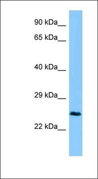
Western Blotting Analysis: Representative lots detected exogenously expressed human and rat CTHRC1 in lysates from transfected cells (Duarte, C.W., et al. (2014). PLoS One.;9(6):e100449; Stohn, J.P., et al. (2012). PLoS One. 7(10):e47142).
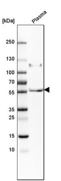
Western Blotting Analysis: A representative lot detected the overexpressed Cthrc1 in plasma samples from Cthrc1 trangenic mice, as well as immunoprecipitated CTHRC1 from normal human plasma samples (Stohn, J.P., et al. (2012). PLoS One. 7(10):e47142).
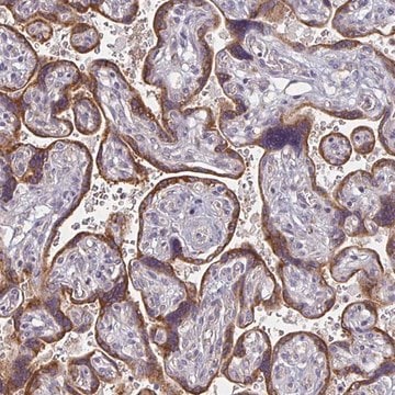
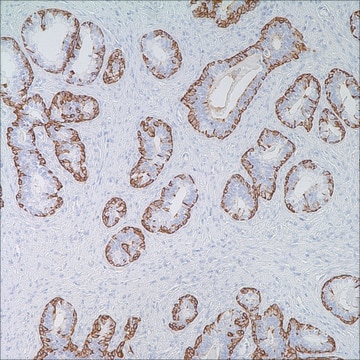
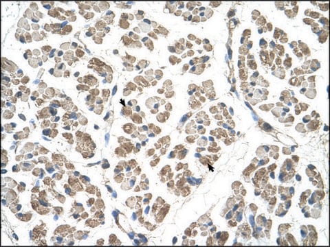
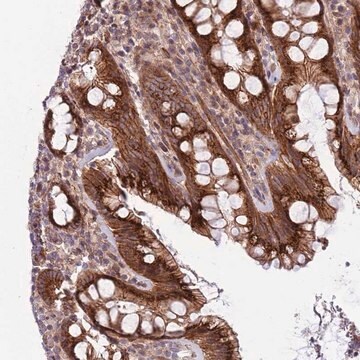
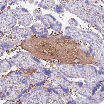
Immunohistochemistry Analysis: A representative lot detected increased Cthrc1 expression in interstitial cells and activated fibroblasts of various remodeling tissues from wild-type, but not Cthrc1-null mice following angiotensin II infusion by immunohistochemistry staining of paraformaldehyde-fixed, paraffin-embedded sections (Duarte, C.W., et al. (2014). PLoS One.;9(6):e100449).
Immunohistochemistry Analysis: Representative lots immunostained Cthrc1-positive cells in paraformaldehyde-fixed, paraffin-embedded human, pig, and rat pituitary gland tissue sections (Duarte, C.W., et al. (2014). PLoS One.;9(6):e100449; Stohn, J.P., et al. (2012). PLoS One. 7(10):e47142).
Immunohistochemistry Analysis: A representative lot immunostained adventitial cells of remodeling renal artery, dermal cells in skin wounds, embryonic cartilage, as well as midbrain of wild-type, but not Cthrc1-null mice, using paraformaldehyde-fixed, paraffin-embedded tissue sections (Stohn, J.P., et al. (2012). PLoS One. 7(10):e47142).
Immunohistochemistry Analysis: A representative lot localized Cthrc1 immunoreactivity in various regions of paraformaldehyde-fixed, paraffin-embedded mouse and pig brain sections (Stohn, J.P., et al. (2012). PLoS One. 7(10):e47142).
Cell Structure
Adhesion (CAMs)
Qualité
Western Blotting Analysis: 10 µg/mL of this antibody detected CTHRC1 in 10 µg of human brain tissue lysate.
Description de la cible
Forme physique
Stockage et stabilité
Autres remarques
Clause de non-responsabilité
Vous ne trouvez pas le bon produit ?
Essayez notre Outil de sélection de produits.
Code de la classe de stockage
12 - Non Combustible Liquids
Classe de danger pour l'eau (WGK)
WGK 1
Point d'éclair (°F)
Not applicable
Point d'éclair (°C)
Not applicable
Certificats d'analyse (COA)
Recherchez un Certificats d'analyse (COA) en saisissant le numéro de lot du produit. Les numéros de lot figurent sur l'étiquette du produit après les mots "Lot" ou "Batch".
Déjà en possession de ce produit ?
Retrouvez la documentation relative aux produits que vous avez récemment achetés dans la Bibliothèque de documents.
Notre équipe de scientifiques dispose d'une expérience dans tous les secteurs de la recherche, notamment en sciences de la vie, science des matériaux, synthèse chimique, chromatographie, analyse et dans de nombreux autres domaines..
Contacter notre Service technique