Accéder au contenu
Merck
Toutes les photos(1)

Key Documents

MAB2262

Sigma-Aldrich

Anti-Excitatory amino acid transporter 2 Antibody, clone G6

clone G6, from mouse

Synonyme(s) :

Glutamate/aspartate transporter II, Sodium-dependent glutamate/aspartate transporter 2, Solute carrier family 1 member 2, excitatory amino acid transporter 2, excitotoxic amino acid transporter 2, glial high affinity glutamate transporter, solute carrier

Se connecterpour consulter vos tarifs contractuels et ceux de votre entreprise/organisme


About This Item

Code UNSPSC :
12352203
eCl@ss :
32160702
Nomenclature NACRES :
NA.41

Source biologique

mouse

Niveau de qualité

Forme d'anticorps

purified immunoglobulin

Type de produit anticorps

primary antibodies

Clone

G6, monoclonal

Espèces réactives

mouse, human

Réactivité de l'espèce (prédite par homologie)

rat (100% immunogen homology)

Technique(s)

immunocytochemistry: suitable
immunohistochemistry: suitable
western blot: suitable

Isotype

IgG1κ

Numéro d'accès NCBI

Numéro d'accès UniProt

Conditions d'expédition

wet ice

Modification post-traductionnelle de la cible

unmodified

Informations sur le gène

human ... SLC1A2(6506)

Description générale

The activity of the main glutamate transporter in the CNS, Excitatory amino acid transporter 2 (also known as GLT1), can be regulated by protein kinase C (PKC). It is known that activation of PKC by phorbol esters promotes the clathrin-dependent internalization of the transporter, followed by its lysosomal degradation. However, the molecular mechanisms that link PKC activation and the internalization of the excitatory amino acid transporter 2 are not fully understood. The internalization process may play a role in the ubiquitylation of lysine residues located in the C-terminal tail of the excitatory amino acid transporter 2. Exposure to PMA increases the ubiquitylation of the excitatory amino acid transporter 2 in transfected cells and in the rat brain, and this ubiquitylated excitatory amino acid transporter 2 accumulates in the intracellular compartment. The activation of PKC induces the ubiquitylation of these C-terminal lysine residues in the Excitatory amino acid transporter 2 and this modification mediates the interaction of the transporter with the endocytic machinery.

Spécificité

This antibody recognizes the C-terminus of Excitatory amino acid transporter 2.

Immunogène

Epitope: C-terminus
Linear peptide corresponding to the C-terminus of human Excitatory amino acid transporter 2.

Application

Research Category
Neuroscience
Research Sub Category
Signaling Neuroscience
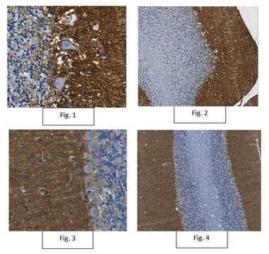
This Anti-Excitatory amino acid transporter 2 Antibody, clone G6 is validated for use in WB, IH, IC for the detection of Excitatory amino acid transporter 2.
Western Blot Analysis: 0.5 µg/ml from a previous lot detected Excitatory amino acid transporter 2 on 10 µg of human brain tissue lysate.

Qualité

Evaluated by Western Blot in mouse brain tissue lysate.

Western Blot Analysis: 0.5 µg/ml of this antibody detected Excitatory amino acid transporter 2 on 10 µg of mouse brain tissue lysate.

Description de la cible

62 kDa

Forme physique

Format: Purified
Protein G Purified
Purified mouse monoclonal IgG1κ in buffer containing 0.1 M Tris-Glycine (pH 7.4, 150 mM NaCl) with 0.05% sodium azide.

Stockage et stabilité

Stable for 1 year at 2-8°C from date of receipt.

Remarque sur l'analyse

Control
Mouse brain tissue lysate

Clause de non-responsabilité

Unless otherwise stated in our catalog or other company documentation accompanying the product(s), our products are intended for research use only and are not to be used for any other purpose, which includes but is not limited to, unauthorized commercial uses, in vitro diagnostic uses, ex vivo or in vivo therapeutic uses or any type of consumption or application to humans or animals.

Vous ne trouvez pas le bon produit ?  

Essayez notre Outil de sélection de produits.

Code de la classe de stockage

12 - Non Combustible Liquids

Classe de danger pour l'eau (WGK)

WGK 1

Point d'éclair (°F)

Not applicable

Point d'éclair (°C)

Not applicable


Certificats d'analyse (COA)

Recherchez un Certificats d'analyse (COA) en saisissant le numéro de lot du produit. Les numéros de lot figurent sur l'étiquette du produit après les mots "Lot" ou "Batch".

Déjà en possession de ce produit ?

Retrouvez la documentation relative aux produits que vous avez récemment achetés dans la Bibliothèque de documents.

Consulter la Bibliothèque de documents

Keodavanh Chounlamountry et al.
Journal of neurochemistry, 134(5), 857-864 (2015-06-18)
Besides the well-described inflammatory and dysfunction effects on the respiratory tract, accumulating evidence indicates that ozone (O3 ) exposure also affects central nervous system functions. However, the mechanisms through which O3 exerts toxic effects on the brain remain poorly understood.
M Asada-Utsugi et al.
Molecular brain, 14(1), 23-23 (2021-01-27)
N-cadherin is a homophilic cell adhesion molecule that stabilizes excitatory synapses, by connecting pre- and post-synaptic termini. Upon NMDA receptor (NMDAR) activation by glutamate, membrane-proximal domains of N-cadherin are cleaved serially by a-disintegrin-and-metalloprotease 10 (ADAM10) and then presenilin 1(PS1, catalytic
Ana Carolina Pinheiro Campos et al.
International journal of molecular sciences, 23(20) (2022-10-28)
Deep brain stimulation (DBS) of the subthalamic nucleus (STN) is considered the gold-standard treatment for PD; however, underlying therapeutic mechanisms need to be comprehensively elucidated, especially in relation to glial cells. We aimed to understand the effects of STN-microlesions and
Michael Ntim et al.
Cerebral cortex (New York, N.Y. : 1991), 30(8), 4617-4632 (2020-03-29)
Synaptic plasticity is the neural basis of physiological processes involved in learning and memory. Tripartite motif-containing 32 (TRIM32) has been found to play many important roles in the brain such as neural stem cell proliferation, neurogenesis, inhibition of nerve proliferation
Lei Qin et al.
Current protocols, 3(12), e964-e964 (2023-12-22)
Astrocytes, the most abundant cells in the central nervous system (CNS), are essential for neuronal development, network formation, and overall CNS homeostasis. Primary astrocyte culture has been successfully used as a tool to study astrocyte biology in vitro. In the

Notre équipe de scientifiques dispose d'une expérience dans tous les secteurs de la recherche, notamment en sciences de la vie, science des matériaux, synthèse chimique, chromatographie, analyse et dans de nombreux autres domaines..

Contacter notre Service technique