Accéder au contenu
Merck
Toutes les photos(1)

Key Documents

16-103

Sigma-Aldrich

Anti-Phosphotyrosine Antibody, clone 4G10®, Biotin Conjugate

clone 4G10®, Upstate®, from mouse

Synonyme(s) :

4G10 Biotin Antibody, Biotinylated Anti-Phosphotyrosine, Clone 4G10 Anti-Phosphotyrosine

Se connecterpour consulter vos tarifs contractuels et ceux de votre entreprise/organisme


About This Item

Code UNSPSC :
12352203
eCl@ss :
32160702
Nomenclature NACRES :
NA.41

Source biologique

mouse

Niveau de qualité

Conjugué

biotin conjugate

Forme d'anticorps

purified antibody

Type de produit anticorps

primary antibodies

Clone

4G10®, monoclonal

Espèces réactives

human

Réactivité de l'espèce (prédite par homologie)

all

Fabricant/nom de marque

Upstate®

Technique(s)

immunocytochemistry: suitable
immunoprecipitation (IP): suitable
western blot: suitable

Isotype

IgG2bκ

Conditions d'expédition

wet ice

Modification post-traductionnelle de la cible

phosphorylation (pTyr)

Informations sur le gène

human ... PID1(55022)

Description générale

Anti-phosphotyrosine monoclonal antibody, clone 4G10 (cat.# 05-321), cross-linked to biotin.
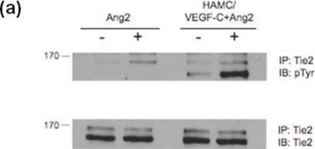
Some of the tyrosine residues can be tagged with a phosphate group (phosphorylated) by protein kinases. (In its phosphorylated state, it is referred to as phosphotyrosine.). Tyrosine phosphorylation is considered as one of the key steps in signal transduction and regulation of enzymatic activity. The advent of anti-phospho-tyrosine antibodies is one of significant events in signal transduction research. Before the availability of anti-phosphotyrosine antibodies, tyrosyl phospyhorylation of proteins and enzymes was investigated through hazardous and time-consuming radioactive experiments. Anti-phosphotyrosine antibodies are commonly used in western blots after the targeted proteins have been immunoprecipitated to measure the tyrosyl phosphorylation of the proteins. Anti-phosphotyrosine antibodies are also directly used on cell lysate to examine the overall change of tyrosine phosphorylation level in response to various treatments.

Description
Anti-phosphotyrosine monoclonal antibody, clone 4G10 (cat. # 05-321), cross-linked to biotin.

Spécificité

Expected to cross-react with rat and mouse.
Recognizes tyrosine-phosphorylated proteins from all species.

Immunogène

Phosphotyramine-KLH.

Application

Anti-Phosphotyrosine Antibody, clone 4G10, Biotin Conjugate detects tyrosine phosphorylated proteins in all species. This unique monoclonal antibody is published in peer reviewed journals and validated for use in IC, IP, WB.
Immunoprecipitation:
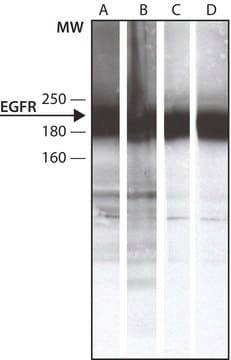
4 μg of a previous lot, used in conjunction with Streptavidin, agarose conjugate (Catalog # 16-126), immunoprecipitated phosphotyrosine containing proteins from a lysate of EGFstimulated A431 cells.
Note: To preserve phosphotyrosine, add 0.2 mM sodium orthovanadate to the lysis buffer.

Immunocytochemistry:
5-10 μg/mL of a previous lot gave positive immunostaining of EGF-stimulated A431 cells that had been fixed with ethanol:acetic acid [1:1].

Application Notes
For use in applications in which a biotin conjugate is advantageous, such as WB and IC.
Research Category
Signaling
Research Sub Category
General Post-translation Modification

Qualité

Routinely evaluated by Western Blot on EGF treated A431 lysates.

Western Blot Analysis:
1:500 dilution of this lot detected Tyrosine phosphorylated proteins on 10 μg of EGF treated A431 lysates.

Description de la cible

Dependent upon the molecular weight of the tyrosine phosphorylated protein being detected.

Forme physique

PBS containing 0.05% sodium azide.
Protein G Purified

Stockage et stabilité

9 months at 2-8°C from date of shipment. NOTE: DO NOT FREEZE. For maximum recovery of product, centrifuge the original vial prior to removing the cap.

Remarque sur l'analyse

Control
Pervanadate-treated human A431 cell extracts or EGF-treated human A431 cells

Autres remarques

Concentration: Please refer to the Certificate of Analysis for the lot-specific concentration.

Informations légales

4G10 is a registered trademark of Upstate Group, Inc.
UPSTATE is a registered trademark of Merck KGaA, Darmstadt, Germany

Clause de non-responsabilité

Unless otherwise stated in our catalog or other company documentation accompanying the product(s), our products are intended for research use only and are not to be used for any other purpose, which includes but is not limited to, unauthorized commercial uses, in vitro diagnostic uses, ex vivo or in vivo therapeutic uses or any type of consumption or application to humans or animals.

Vous ne trouvez pas le bon produit ?  

Essayez notre Outil de sélection de produits.

Code de la classe de stockage

12 - Non Combustible Liquids

Classe de danger pour l'eau (WGK)

WGK 2

Point d'éclair (°F)

Not applicable

Point d'éclair (°C)

Not applicable


Certificats d'analyse (COA)

Recherchez un Certificats d'analyse (COA) en saisissant le numéro de lot du produit. Les numéros de lot figurent sur l'étiquette du produit après les mots "Lot" ou "Batch".

Déjà en possession de ce produit ?

Retrouvez la documentation relative aux produits que vous avez récemment achetés dans la Bibliothèque de documents.

Consulter la Bibliothèque de documents

Occurrence and phase distribution of selected pharmaceuticals in the Yangtze Estuary and its coastal zone.
Yang Y, Fu J, Peng H, Hou L, Liu M, Zhou JL
Journal of Hazardous Materials null
Christopher N Davis et al.
Proceedings of the National Academy of Sciences of the United States of America, 103(8), 2953-2958 (2006-02-16)
Interleukin (IL)-1beta is a pluripotent proinflammatory cytokine that signals through the type-I IL-1 receptor (IL-1RI), a member of the Toll-like receptor family. In hypothalamic neurons, binding of IL-1beta to IL-1RI mediates transcription-dependent changes that depend on the recruitment of the
W H Ho et al.
The Journal of biological chemistry, 270(24), 14523-14532 (1995-06-16)
The heregulin family of polypeptides arise as splice variants from a single gene and share a conserved epidermal growth factor (EGF)-like domain thought to be the major determinant of their biological activities. We report here the cloning of a novel
Imran H Khan et al.
Molecular & cellular proteomics : MCP, 5(4), 758-768 (2005-12-22)
Phosphorylation analysis of signaling proteins is key for examining intracellular signaling pathways. Conventional biochemical approaches, e.g. immunoprecipitation, Western blot, and ELISA, have played a major role in elucidation of individual signaling events. However, these methods are laborious, time-consuming, and difficult
H Nishizumi et al.
The Journal of experimental medicine, 187(8), 1343-1348 (1998-05-23)
B cells from young lyn-/- mice are hyperresponsive to anti-IgM-induced proliferation, suggesting involvement of Lyn in negative regulation of B cell antigen receptor (BCR)-mediated signaling. Here we show that tyrosine phosphorylation of FcgammaRIIB and CD22 coreceptors, which are important for

Notre équipe de scientifiques dispose d'une expérience dans tous les secteurs de la recherche, notamment en sciences de la vie, science des matériaux, synthèse chimique, chromatographie, analyse et dans de nombreux autres domaines..

Contacter notre Service technique