Saltar al contenido
MilliporeSigma
Todas las fotos(4)

Documentos

MABT1340

Sigma-Aldrich

Anti-Lamin A/C Antibody, clone 2A1

clone 2A1, from mouse

Sinónimos:

70 kDa Lamin, Renal carcinoma antigen NY-REN-32

Iniciar sesiónpara Ver la Fijación de precios por contrato y de la organización


About This Item

UNSPSC Code:
12352203
eCl@ss:
32160702
NACRES:
NA.41

biological source

mouse

antibody form

purified immunoglobulin

antibody product type

primary antibodies

clone

2A1, monoclonal

species reactivity

mouse, monkey, hamster, human

packaging

antibody small pack of 25 μg

technique(s)

immunocytochemistry: suitable
immunofluorescence: suitable
immunoprecipitation (IP): suitable
western blot: suitable

isotype

IgG2bκ

NCBI accession no.

UniProt accession no.

target post-translational modification

unmodified

Gene Information

human ... LMNA(4000)

General description

Prelamin-A/C (UniProt: P02545) is encoded by the LMNA (also known as LMN1) gene (Gene ID: 4000) in human. It is cleaved into Lamin-A/C (also known as 70 kDa Lamin, Renal carcinoma antigen NY-REN-32). Lamins are components of the nuclear lamina that provide a framework for the nuclear envelope and may also interact with chromatin. Lamin A and C are present in equal amounts in the lamina of mammals. Plays an important role in nuclear assembly, chromatin organization, nuclear membrane and telomere dynamics. Lamin A is initially synthesized as prelamin A that undergoes several modifications in the carboxyl terminal region that allow incorporation of prelamin A into the nuclear envelope and its subsequent processing into the mature lamin A. Cleavage of 15 residues (aa 647-662) by ZMPSTE24/FACE1 generates the final protein product. Unlike mature lamin A, prelamin A accumulates as discrete and localized foci at the nuclear periphery. Prelamin-A/C can accelerate smooth muscle cell senescence. It can act to disrupt mitosis and induce DNA damage in vascular smooth muscle cells (VSMCs), leading to mitotic failure, genomic instability, and premature senescence. Mutations in LMNA gene are known to cause Emery-Dreifuss muscular dystrophy that is characterized by weakness and atrophy of muscle without involvement of the nervous system. Some mutations have also been linked to familial type of lipodystrophy characterized by the loss of subcutaneous adipose tissue in the lower parts of the body. (Ref.: Casasola, A., et al. (2016). Nucleus 7(1); 84-102).

Specificity

Clone 2A1 detects the Lamin A/C species in the nuclear interior in multiple species. It targets the Rod domain of Lamin A/C.

Immunogen

Purified protein corresponding to the rod domain for human Lamin A/C.

Application

Anti-Lamin A/C, clone 2A1, Cat. No. MABT1340, is a highly specific mouse monoclonal antibody that targets Lamin A/C and has been tested for use in Immunocytochemistry, Immunofluorescence, Immunoprecipitation, and Western Blotting.
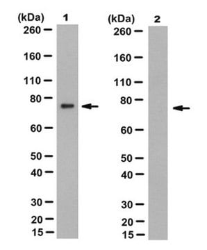
Immunoprecipitation Analysis: A representative immunoprecipitated Lamin A/C in culture supernatant (Data courtesy of Marie Lang, M.D., Stefan Schuchner, Ph.D. and Egon Ogris, M.D., Medical University of Vienna, Austria).


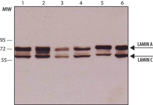
Western Blotting Analysis: A representative lot detected Lamin A/C in culture supernatants of various cell lines (Data courtesy of Marie Lang, M.D., Stefan Schuchner, Ph.D. and Egon Ogris, M.D., Medical University of Vienna, Austria).


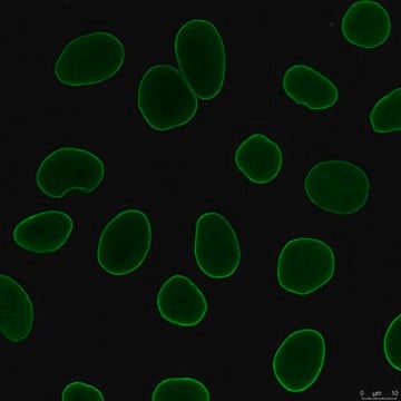
Immunocytochemistry Analysis: A 1:1,000 dilution from a representative lot detected Lamin A/C in HeLa cells.


Immunofluorescence Analysis: A representative lot detected Lamin A/C in the nuclear interior of HeLa cells (Data courtesy of Marie Lang, M.D., Stefan Schuchner, Ph.D. and Egon Ogris, M.D., Medical University of Vienna, Austria).
Research Category
Cell Structure

Quality

Evaluated by Western Blotting in HeLa cell lysate.

Western Blotting Analysis: 0.2 µg/mL of this antibody detected Lamin A/C in HeLa cell lysate.

Target description

74 and 65 kDa observed. 74.14 kDa and 65.14 kDa for Lamin A and C. Uncharacterized bands may be observed in some lysate(s).

Physical form

Format: Purified
Protein G purified
Purified mouse monoclonal antibody IgG2b in buffer containing 0.1 M Tris-Glycine (pH 7.4), 150 mM NaCl with 0.05% sodium azide.

Storage and Stability

Stable for 1 year at 2-8°C from date of receipt.

Other Notes

Concentration: Please refer to lot specific datasheet.

Disclaimer

Unless otherwise stated in our catalog or other company documentation accompanying the product(s), our products are intended for research use only and are not to be used for any other purpose, which includes but is not limited to, unauthorized commercial uses, in vitro diagnostic uses, ex vivo or in vivo therapeutic uses or any type of consumption or application to humans or animals.

Not finding the right product?  

Try our Herramienta de selección de productos.


Certificados de análisis (COA)

Busque Certificados de análisis (COA) introduciendo el número de lote del producto. Los números de lote se encuentran en la etiqueta del producto después de las palabras «Lot» o «Batch»

¿Ya tiene este producto?

Encuentre la documentación para los productos que ha comprado recientemente en la Biblioteca de documentos.

Visite la Librería de documentos

Tommaso Cavazza et al.
Cell, 184(11), 2860-2877 (2021-05-09)
Most human embryos are aneuploid. Aneuploidy frequently arises during the early mitotic divisions of the embryo, but its origin remains elusive. Human zygotes that cluster their nucleoli at the pronuclear interface are thought to be more likely to develop into

Nuestro equipo de científicos tiene experiencia en todas las áreas de investigación: Ciencias de la vida, Ciencia de los materiales, Síntesis química, Cromatografía, Analítica y muchas otras.

Póngase en contacto con el Servicio técnico