Skip to Content
MilliporeSigma
All Photos(2)

Documents

AMAB90569

Sigma-Aldrich

Monoclonal Anti-NLRP3 antibody produced in mouse

Prestige Antibodies® Powered by Atlas Antibodies, clone CL0210, purified immunoglobulin, buffered aqueous glycerol solution

Synonym(s):

Anti-AGTAVPRL, Anti-AII, Anti-AVP, Anti-C1orf7, Anti-CIAS1, Anti-CLR1.1, Anti-FCAS, Anti-FCU, Anti-MWS, Anti-NALP3, Anti-PYPAF1

Sign Into View Organizational & Contract Pricing


About This Item

UNSPSC Code:
12352203
NACRES:
NA.43

biological source

mouse

Quality Level

conjugate

unconjugated

antibody form

purified immunoglobulin

antibody product type

primary antibodies

clone

CL0210, monoclonal

product line

Prestige Antibodies® Powered by Atlas Antibodies

form

buffered aqueous glycerol solution

species reactivity

human

technique(s)

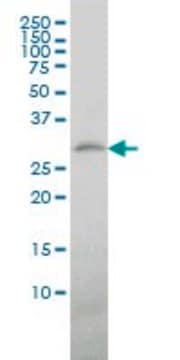
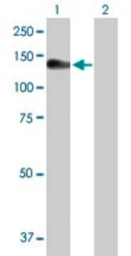
immunoblotting: 1 μg/mL
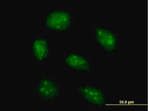
immunofluorescence: 2-10 μg/mL (Fixation/Permeabilization: PFA/Triton X-100)

isotype

IgG1

immunogen sequence

FKMHLEDYPPQKGCIPLPRGQTEKADHVDLATLMIDFNGEEKAWAMAVWIFAAINRRDLYEKAKRDEPKWGSDNARVSNPTVICQEDSIEEEWMGLLEYLSRISICKMKKDYRKKYR

UniProt accession no.

shipped in

wet ice

storage temp.

−20°C

target post-translational modification

unmodified

Gene Information

human ... NLRP3(114548)

Immunogen

NLR family, pyrin domain containing 3

Application

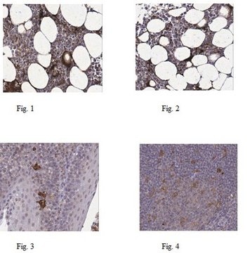
All Prestige Antibodies® Powered by Atlas Antibodies is developed and validated by the Human Protein Atlas (HPA) project . Each antibody is tested by immunohistochemistry against hundreds of normal and disease tissues. These images can be viewed on the Human Protein Atlas (HPA) site by clicking on the Image Gallery link. We also provide Prestige Antibodies® protocols and other useful information.

Features and Benefits

Prestige Antibodies® are highly characterized and extensively validated antibodies with the added benefit of all available characterization data for each target being accessible via the Human Protein Atlas portal linked just below the product name at the top of this page. The uniqueness and low cross-reactivity of the Prestige Antibodies® to other proteins are due to a thorough selection of antigen regions, affinity purification, and stringent selection. Prestige antigen controls are available for every corresponding Prestige Antibody and can be found in the linkage section.

Every Prestige Antibody is tested in the following ways:
  • IHC tissue array of 44 normal human tissues and 20 of the most common cancer type tissues.
  • Protein array of 364 human recombinant protein fragments.

Linkage

Corresponding Antigen APREST81743

Physical form

Phospate buffered saline, pH 7.2, containing 40% glycerol and 0.02% sodium azide

Legal Information

Prestige Antibodies is a registered trademark of Merck KGaA, Darmstadt, Germany

Not finding the right product?  

Try our Product Selector Tool.

Storage Class

10 - Combustible liquids

wgk_germany

WGK 1

flash_point_f

Not applicable

flash_point_c

Not applicable


Certificates of Analysis (COA)

Search for Certificates of Analysis (COA) by entering the products Lot/Batch Number. Lot and Batch Numbers can be found on a product’s label following the words ‘Lot’ or ‘Batch’.

Already Own This Product?

Find documentation for the products that you have recently purchased in the Document Library.

Visit the Document Library

Bing Zhou et al.
Oxidative medicine and cellular longevity, 2020, 6384803-6384803 (2020-06-09)
Vascular oxidative stress and inflammation play a major role in vascular diseases. This study was aimed at determining the protective roles of fibronectin type III domain-containing 5 (FNDC5) in angiotensin II- (Ang II-) induced vascular oxidative stress and inflammation and
Viviane Dias Faustino et al.
Bioscience reports, 38(4) (2018-06-20)
Protein overload of proximal tubular cells (PTCs) can promote interstitial injury by unclear mechanisms that may involve activation of innate immunity. We investigated whether prolonged exposure of tubular cells to high protein concentrations stimulates innate immunity, triggering progressive interstitial inflammation
Roberto Romero et al.
American journal of reproductive immunology (New York, N.Y. : 1989), 79(6), e12440-e12440 (2016-03-10)
Inflammasomes are signaling platforms that, upon sensing pathogens and sterile stressors, mediate the release of mature forms of interleukin (IL)-1β and IL-18. The aims of this study were to determine (i) the expression of major inflammasome components in the chorioamniotic

Our team of scientists has experience in all areas of research including Life Science, Material Science, Chemical Synthesis, Chromatography, Analytical and many others.

Contact Technical Service