推薦產品
生物源
rabbit
品質等級
抗體表格
affinity purified immunoglobulin
抗體產品種類
primary antibodies
無性繁殖
polyclonal
純化經由
affinity chromatography
物種活性
mouse, rat
製造商/商標名
Chemicon®
技術
immunohistochemistry: suitable
immunoprecipitation (IP): suitable
western blot: suitable
NCBI登錄號
UniProt登錄號
運輸包裝
dry ice
目標翻譯後修改
unmodified
基因資訊
mouse ... Kcnc1(16502)
特異性
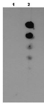
Recognizes a full length Kv3.1b protein. Does not cross react with any other potassium channel antigens tested so far.
免疫原
Purified peptide from rat Kv3.1b (amino acids 567-585) (Accession P25122).
應用
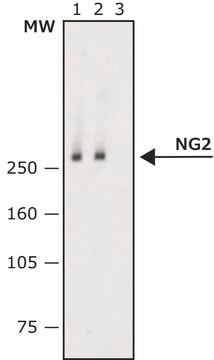
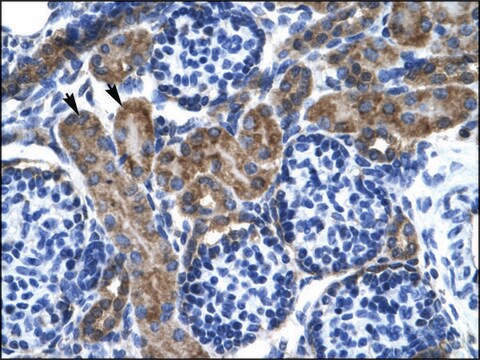
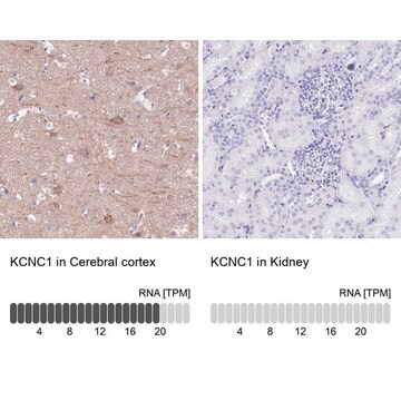
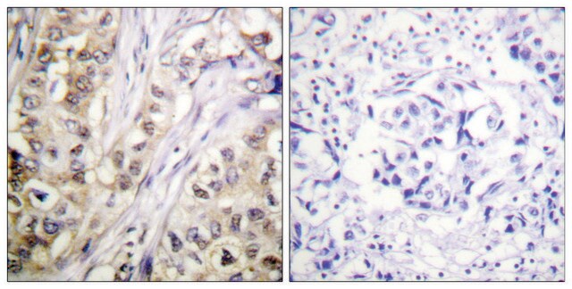
This Anti-Potassium Channel Kv3.1b Antibody is validated for use in IH, IP, WB for the detection of Potassium Channel Kv3.1b.
Western blot: 1:200 using ECL on rat brain membranes.
Immunohistochemistry on rat brain sections.
Immunoprecipitation
Dilutions should be made using a carrier protein such as BSA (1-3%)
Optimal working dilutions must be determined by the end user.
PROTOCOL FOR IMMUNOHISTOCHEMISTRY
Sacrifice and tissue processing:
Rats are deeply anesthetized with pentobarbital sodium (Pental). Brain is fixed by transcardial perfusion, first with 50 mL of phosphate buffered saline (0.02M PBS, pH 7.4) containing heparin (5U/ml), then with 220 mL of ice-cold 4% paraformaldehyde in 0.1M PBS, pH 7.5 containing sucrose 4%. Brain is cut in coronal blocks and further fixed by immersion in the same fixative, refrigerated, for 1-2 hours. Brain blocks are transferred to 10% sucrose in 0.1M PBS and sectioned in a cryostat within 3 days. Brain sections, 30 μm thick, are floated in 0.1M PBS and then preserved in a cryopreservation buffer at -20°C. The cryopreservation buffer contains 40% ethylene glycol and 1% polyvinylpyrrolidone in 0.1M potassium acetate buffer, pH 6.5.
Staining procedure:
1. Floating sections are rinsed in 0.02M PBS, 2x 5 minutes.
2. Endogenous peroxidase activity is quenched by incubation with 0.2 % hydrogen peroxide in 0.1M phosphate buffer pH 7.3 containing 0.2% Triton X-100 for 25 minutes at room temperature.
3. Sections are rinsed in 0.02M PBS, 2x 5 minutes.
4. Sections are incubated with the primary antiserum in a medium containing 0.3% Triton X-100, 0.05% Tween 20, 4% normal donkey serum (NDS), for 1 hour at room temperature and then overnight refrigerated.
5. Sections are rinsed in 0.02M PBS, containing 4% NDS, 2x 5 minutes.
Sections are incubated with biotinylated donkey anti-rabbit (from Chemicon USA, catalog number AP182B) diluted 1:400 in 0.02M PBS, containing 0.3% Triton X-100, 0.05% Tween 20, and 4% NDS, for 1 hour at room temperature and then overnight refrigerated.
1. Sections are rinsed in 0.02M PBS containing 4% NDS, 2x 5 min.
2. Sections are incubated with extravidin-peroxidase (Sigma Catalog number E2886) diluted 1:100 in 0.02M PBS, for 45 minutes at room temperature.
3. Sections are rinsed in 0.02M PBS, 3x 5 minutes.
Color development:
1. Sections are incubated with a solution of diaminobenzidine at the concentration of 0.0125% and containing 0.05% nickel ammonium sulfate for 10 minutes at room temperature.
2. Sections are transferred to the same DAB solution but with added hydrogen peroxide at a final concentration of 0.0015%. Duration of incubation should be adjusted by the end user.
3. Sections are rinsed in 0.02M PBS, 4x 10 minutes.
4. Sections are mounted on glass slides (gelatinized or coated by any other type of adhesive material) and allowed to dry.
5. Sections are dehydrated in ascending series of ethanol concentrations (70%, 90%, 100%, 5 minutes in each), delipidated in xylene (10 minutes) and coverslipped in Permount (or any other xylene diluted adhesive).
分析報告
Control
CONTROL ANTIGEN: Included free of charge with the antibody is 40 μg of control antigen (lyophilized powder in phosphate buffered saline, pH 7.4, containing 5% sucrose and 0.025% sodium azide). The stock solution of the antigen can be made up using 200 μL of sterile deionized water. For negative control, preincubate 1 μg of peptide with 1 μg of antibody for one hour at room temperature. Optimal concentrations must be determined by the end user.
CONTROL ANTIGEN: Included free of charge with the antibody is 40 μg of control antigen (lyophilized powder in phosphate buffered saline, pH 7.4, containing 5% sucrose and 0.025% sodium azide). The stock solution of the antigen can be made up using 200 μL of sterile deionized water. For negative control, preincubate 1 μg of peptide with 1 μg of antibody for one hour at room temperature. Optimal concentrations must be determined by the end user.
其他說明
Concentration: Please refer to the Certificate of Analysis for the lot-specific concentration.
法律資訊
CHEMICON is a registered trademark of Merck KGaA, Darmstadt, Germany
Not finding the right product?
Try our 產品選擇工具.
儲存類別代碼
11 - Combustible Solids
水污染物質分類(WGK)
WGK 3
分析證明 (COA)
輸入產品批次/批號來搜索 分析證明 (COA)。在產品’s標籤上找到批次和批號,寫有 ‘Lot’或‘Batch’.。
David Soares et al.
The Journal of comparative neurology, 525(9), 2164-2174 (2017-02-19)
There are substantial differences across species in the organization and function of the motor pathways. These differences extend to basic electrophysiological properties. Thus, in rat motor cortex, pyramidal cells have long duration action potentials, while in the macaque, some pyramidal
Hui Hong et al.
Frontiers in cellular neuroscience, 12, 175-175 (2018-07-13)
In the auditory system, tonotopy is the spatial arrangement of where sounds of different frequencies are processed. Defined by the organization of neurons and their inputs, tonotopy emphasizes distinctions in neuronal structure and function across topographic gradients and is a
Dean O Smith et al.
PloS one, 3(2), e1604-e1604 (2008-02-14)
Because of the importance of voltage-activated K(+) channels during embryonic development and in cell proliferation, we present here the first description of these channels in E15 rat embryonic neural progenitor cells derived from the subventricular zone (SVZ). Activation, inactivation, and
我們的科學家團隊在所有研究領域都有豐富的經驗,包括生命科學、材料科學、化學合成、色譜、分析等.
聯絡技術服務

