Skip to Content
Merck
All Photos(4)

Key Documents

MAB4190

Sigma-Aldrich

Anti-Ki-67 Antibody, clone Ki-S5

clone Ki-S5, Chemicon®, from mouse

Sign Into View Organizational & Contract Pricing


About This Item

UNSPSC Code:
12352203
eCl@ss:
32160702
NACRES:
NA.41

biological source

mouse

Quality Level

antibody form

purified immunoglobulin

antibody product type

primary antibodies

clone

Ki-S5, monoclonal

species reactivity

human

packaging

antibody small pack of 25 μg

manufacturer/tradename

Chemicon®

technique(s)

flow cytometry: suitable
immunocytochemistry: suitable
immunohistochemistry (formalin-fixed, paraffin-embedded sections): suitable
western blot: suitable

isotype

IgG1

NCBI accession no.

UniProt accession no.

shipped in

ambient

storage temp.

2-8°C

target post-translational modification

unmodified

Gene Information

human ... MKI67(4288)

General description

Ki67 antigen is the prototypic cell cycle related nuclear protein, expressed by proliferating cells in all phases of the active cell cycle (G1, S, G2 and M phase). It is absent in resting (G0) cells. Ki67 antibodies are useful in establishing the cell growing fraction in neoplasms (immunohistochemically quantified by determining the number of Ki67 positive cells among the total number of resting cells = Ki67 index). In neoplastic tissues the prognostic value is comparable to the tritiated thymidine labelling index. The correlation between low Ki67 index and histologically low grade tumours is strong. Ki67 is routinely used as a marker of cell cycling and proliferation. This specificity of Ki-S5 antibody for proliferating cells might make it a useful tool for determination of the proliferative fraction in tumors such as Non-Hodgkin′s lymphoma (Kreipe et al., 1993), mammary carcinomas (Mauri et al., 1984), ovarian tumors (Ballin et al., 1994) and prostate cancer.

Specificity

In Immunoprecipitation and Western blot experiments the Ki-S5 antibody recognizes a cell-cycle-associated protein of 345 kD and 395 kD (Kreipe et al., 1993) identical with the Ki-67 antigen. The antibody binds to a formalin-resistant epitope of the Ki-67 antigen. The immunoreactivity of Ki-S5 is confined to the nuclei of proliferating cells and no crossreactivity with cytoplasmic antigens of epithelial occurs even after antigen retrieval (Mauri et al., 1984). A comparison of immunohistochemical labeling of fresh and fixed tissue samples of NHL showed that identical results were obtained with Ki-67 and Ki-S5 (Kreipe et al., 1993). The Ki-67 antigen is preferentially expressed during late G1, S, G2 and M phase of the cell cycle, while resting, non-cycling cells (G0 phase) lack the Ki-67 antigen. In addition, constantly proliferating cells (e.g. cell lines) react positively to Ki-S5 during the entire cell-cycle. This specificity of Ki-S5 antibody for proliferating cells might make it a useful tool for determination of the proliferative fraction in tumors such as Non-Hodgkin′s lymphoma (Kreipe et al., 1993), mammary carcinomas (Mauri et al., 1984), ovarian tumors (Ballin et al., 1994) and prostate cancer.

Immunogen

Nuclear protein preparation from the human cell line U937.50.

Application

Anti-Ki-67 Antibody, clone Ki-S5 is a high quality Mouse Monoclonal Antibody for the detection of Ki-67 & has been validated in FC, ICC, IHC, IHC(P) & WB.
Immunocytochemistry:
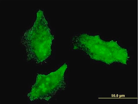
A 5-10 μg/mL concentration of a previous lot was used in IC.

Immunohistochemistry:
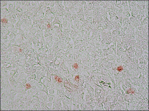
A 5-10 μg/mL concentration of a previous lot was used in IH.

Immunohistochemistry:
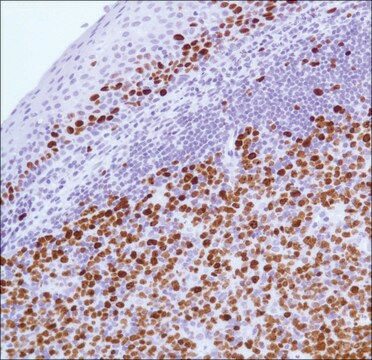
5-10 µg/mL on formalin fixed paraffin tissue. Citrate buffer-microwave antigen retrieval

Flow Cytometry:
A previous lot of this antibody was used in FC.

Western blot:
1-10 µg/mL

Optimal working dilutions must be determined by end user.
Research Category
Epigenetics & Nuclear Function
Research Sub Category
Cell Cycle, DNA Replication & Repair

Quality

Evaluated by Western Blot on A431 lysates.

Western Blot Analysis:
1:500 dilution of this antibody detected cell-cycle-associated protein of 345 kDa and 395 kDa on 10 μg of A431 lysates.

Target description

345 & 395 kDa

Physical form

Format: Purified
Mouse monoclonal IgG1 in buffer containing 0.02 M Phosphate buffer, 0.25 M NaCl, pH 7.6 with 0.1% sodium azide.
Protein A purified

Storage and Stability

Stable for 1 year at 2-8ºC from date of receipt.

Analysis Note

Control
Tonsil tissue, A431 cell lysate

Other Notes

Concentration: Please refer to the Certificate of Analysis for the lot-specific concentration.

Legal Information

CHEMICON is a registered trademark of Merck KGaA, Darmstadt, Germany

Disclaimer

Unless otherwise stated in our catalog or other company documentation accompanying the product(s), our products are intended for research use only and are not to be used for any other purpose, which includes but is not limited to, unauthorized commercial uses, in vitro diagnostic uses, ex vivo or in vivo therapeutic uses or any type of consumption or application to humans or animals.

Not finding the right product?  

Try our Product Selector Tool.

Storage Class Code

10 - Combustible liquids

WGK

WGK 2

Flash Point(F)

Not applicable

Flash Point(C)

Not applicable


Certificates of Analysis (COA)

Search for Certificates of Analysis (COA) by entering the products Lot/Batch Number. Lot and Batch Numbers can be found on a product’s label following the words ‘Lot’ or ‘Batch’.

Already Own This Product?

Find documentation for the products that you have recently purchased in the Document Library.

Visit the Document Library

Paula Rocktäschel et al.
Epilepsy & behavior : E&B, 101(Pt B), 106581-106581 (2019-11-26)
Tuberous sclerosis complex (TSC) is a neurodevelopmental disorder caused by deletions in the TSC1 or TSC2 genes that is associated with epilepsy in up to 90% of patients. Seizures are suggested to start in benign brain tumors, cortical tubers, or
Sandra Haider et al.
Endocrinology, 155(1), 263-274 (2013-11-06)
Failures in human extravillous trophoblast (EVT) development could be involved in the pathogenesis of pregnancy diseases. However, the underlying mechanisms have been poorly characterized. Here, we provide evidence that Notch signaling could represent a key regulatory pathway controlling trophoblast proliferation
Identification of a Src tyrosine kinase/SIAH2 E3 ubiquitin ligase pathway that regulates C/EBP? expression and contributes to transformation of breast tumor cells.
Sarkar, TR; Sharan, S; Wang, J; Pawar, SA; Cantwell, CA; Johnson, PF; Morrison, DK; Wang et al.
Molecular and cellular biology null
Saskia Grudzenski et al.
Stem cell research & therapy, 8(1), 96-96 (2017-04-28)
In the field of experimental stem cell therapy, intra-arterial (IA) delivery yields the best results concerning, for example, migrated cell number at the targeted site. However, IA application also appears to be associated with increased mortality rates and infarction. Since
Sophie Blondel et al.
Stem cells translational medicine, 3(4), 510-519 (2014-03-07)
Hutchinson-Gilford progeria syndrome is a rare congenital disease characterized by premature aging in children. Identification of the mutation and related molecular mechanisms has rapidly led to independent clinical trials testing different marketed drugs with a preclinically documented impact on those

Our team of scientists has experience in all areas of research including Life Science, Material Science, Chemical Synthesis, Chromatography, Analytical and many others.

Contact Technical Service