Skip to Content
Merck
All Photos(3)

Key Documents

MABE415

Sigma-Aldrich

Anti-FoxP2 Antibody, clone FOXP2-73A/8

clone FOXP2-73A/8, from mouse

Synonym(s):

Forkhead box protein P2, CAG repeat protein 44, Trinucleotide repeat-containing gene 10 protein

Sign Into View Organizational & Contract Pricing


About This Item

UNSPSC Code:
12352203
eCl@ss:
32160702
NACRES:
NA.41

biological source

mouse

Quality Level

antibody form

purified immunoglobulin

antibody product type

primary antibodies

clone

FOXP2-73A/8, monoclonal

species reactivity

human

technique(s)

immunohistochemistry: suitable
immunoprecipitation (IP): suitable
western blot: suitable

isotype

IgG1κ

NCBI accession no.

UniProt accession no.

shipped in

wet ice

target post-translational modification

unmodified

Gene Information

human ... FOXP2(93986)

General description

Forkhead box protein P2 (FoxP2) is a multidomain transcriptional repressor that belongs to the Fox family of proteins. The FoxP2 protein contains a forkhead-box DNA-binding domain, characteristic of the FOX group of transcription factors. In addition, this protein contains a polyglutamine tract, as well as zinc finger and leucine zipper motifs. In order for FoxP2 to bind DNA, it forms homodimers and heterodimers with FoxP1 and FoxP4. The forkhead box family of proteins play an important role in immune response, organ development and the development of cancer. Functional studies in vertebrates suggest this gene plays a role in the modulation of neural plasticity. In line with these findings, mutations of FoxP2 in humans have been linked to speech and language disorders.

Specificity

This antibody recognizes FoxP2 isoforms at 70 kDa, 48 kDa, and 40 kDa.

Immunogen

GST-tagged recombinant protein corresponding to the N-terminus of Human FoxP2.

Application

Detect FOXP2 using this mouse monoclonal antibody, Anti-FoxP2 Antibody, clone FOXP2-73A/8 validated for use in western blotting & IHC.
Western Blotting Analysis: 1 µg/mL from a representative lot detected FoxP2 in 10 µg of THP-1 cell lysate.

Immunoprecipitation Analysis (IP): A representative lot of this antibody was demonstrated to immunoprecipitate FoxP2 using growth arrested 143B cells (Gascoyne D.M., et al. (2015). PLoS One, 10(6):e0128513).

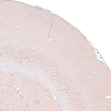
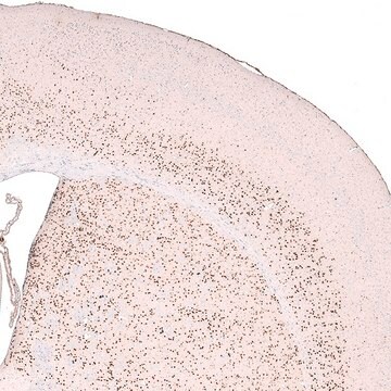
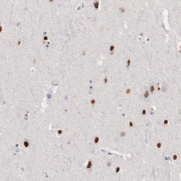
Immunohistochemistry Analysis: A representative lot from an independent laboratory detected FoxP2 in human tonsil tissues (Campbell, A. J., et al. (2010). Br J Haematol. 149(2):221-230.).

Immunohistochemistry Analysis: A representative lot from an independent laboratory detected FoxP2 in proliferating chondrocytes of murine E17.5 long bone (Gascoyne D.M., et al. (2015). PLoS One, 10(6):e0128513).

Quality

Evaluated by Western Blotting in HeLa cell lysate.

Western Blotting Analysis: 1 µg/mL of this antibody detected FoxP2 in 10 µg of HeLa cell lysate.

Target description

~80 kDa observed. Uniprot describes 9 isoforms between 9-83 kDa

Physical form

Format: Purified

Other Notes

Concentration: Please refer to the Certificate of Analysis for the lot-specific concentration.

Not finding the right product?  

Try our Product Selector Tool.

Storage Class Code

12 - Non Combustible Liquids

WGK

WGK 1

Flash Point(F)

Not applicable

Flash Point(C)

Not applicable


Certificates of Analysis (COA)

Search for Certificates of Analysis (COA) by entering the products Lot/Batch Number. Lot and Batch Numbers can be found on a product’s label following the words ‘Lot’ or ‘Batch’.

Already Own This Product?

Find documentation for the products that you have recently purchased in the Document Library.

Visit the Document Library

Pedro M Rodenas-Cuadrado et al.
The Journal of comparative neurology, 526(8), 1235-1266 (2018-01-04)
Genes including FOXP2, FOXP1, and CNTNAP2, have been implicated in human speech and language phenotypes, pointing to a role in the development of normal language-related circuitry in the brain. Although speech and language are unique to humans a comparative approach
Takako Kita et al.
The European journal of neuroscience, 44(11), 2899-2908 (2016-10-08)
Projections from the posterior intralaminar thalamic nuclei and the superior colliculus (SC) to the subthalamic nucleus (STN) and the zona incerta (ZI) have been described in the primate and rodent. The aims of this study was to investigate several questions
Ayla Aksoy-Aksel et al.
eLife, 10 (2021-05-25)
Dopaminergic signaling plays an important role in associative learning, including fear and extinction learning. Dopaminergic midbrain neurons encode prediction error-like signals when threats differ from expectations. Within the amygdala, GABAergic intercalated cell (ITC) clusters receive one of the densest dopaminergic
Boglárka Barsy et al.
Nature neuroscience, 23(5), 625-637 (2020-04-15)
Decades of research support the idea that associations between a conditioned stimulus (CS) and an unconditioned stimulus (US) are encoded in the lateral amygdala (LA) during fear learning. However, direct proof for the sources of CS and US information is
Shane P D'Souza et al.
The Journal of comparative neurology, 530(8), 1247-1262 (2021-11-08)
Obtaining a parts list of the sensory components of the retina is vital to understand the effects of light in behavior, health, and disease. Rods, cones, and intrinsically photosensitive retinal ganglion cells (ipRGCs) are the best described photoreceptors in the

Our team of scientists has experience in all areas of research including Life Science, Material Science, Chemical Synthesis, Chromatography, Analytical and many others.

Contact Technical Service