Poor Cell Growth Troubleshooting
Cell Growth: An Overview
Ensuring adequate cell growth is a critical part of collecting accurate data with cell cultures. Cells can be cultured in suspension, or as a monolayer that attaches to cultureware, such as a flask, dish or multiwell plate. The culture method is determined by the cells’ endogenous phenotype and tissue of origin — so, cells derived from blood generally grow in suspension, while those derived from solid tissues typically grow in monolayers. Co-culturing a mixed population that contains both attached cells as well as phenotypes in suspension is also possible.

Figure 1.Phases of Cell Growth
For cells that grow as a monolayer, confluence is defined as the percentage of the culture vessel surface area that appears covered by a layer of cells when observed by microscopy. For example, 50% confluency means half of the surface of the culture dish, flask, etc. is covered in cells. For suspension cells, growth progress is generally measured by recommended cell density for the line or type – but ‘confluency’ may be assessed by increasing turbidity and stable medium color.
Cell growth in culture generally occurs in four phases that, when graphed as the log of the cell count over time, generates a sigmoidal (S-shaped) curve, although the amount of time spent in each phase differs between individual cell lines and cultures.
Lag Phase – Cells are acclimating to culture conditions and do not divide during this phase. Cell attachment generally occurs within 24 hours after initiation of the culture. The total length of this phase is determined by the growth phase and seeding density of the cells that were used to start the culture.
Log (Logarithmic) Growth Phase – Cells are actively dividing during this phase, and this is the best time for assessing population growth as well as for general data collection. Late in the log phase is the best time to passage (subculture) cells, before overcrowding can lead to cell stress.
Plateau (Stationary) Phase – Cell growth in this phase slows as cells approach 100% confluence, and fewer than one tenth of cells are in the active cell cycle. Cells are most susceptible to injury in this phase; careful observation is therefore needed to ensure cells are passaged prior to or at the outset of this phase.
Decline Phase – As a natural part of the cell cycle, the population of live cells declines as cell death predominates in this phase.
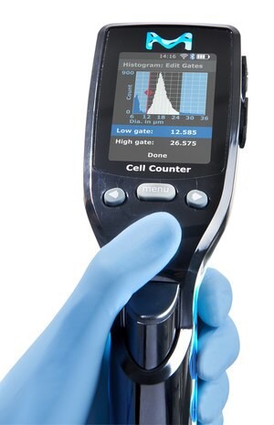
Accurate cell counting is of paramount importance when assessing cell growth in cultures, as faulty counts can lead to mistaken conclusions about cell health. Cell counts can be determined using a hemocytometer. However, automated cell counters that utilize the Coulter principle where cells flow, one by one, through an aperture within an electrical sensor, yield the most precise cell counts.

Figure 2.Scepter™ 3.0 Handheld Automated Cell Counter.
General tips and techniques for preventing and eliminating cell growth problems
In addition to following good sterility practices to prevent microbial and chemical contamination, the keys to early diagnosis and treatment of cell growth problems are frequent observation of cultures and detailed recordkeeping. It’s also important to consider whether the most efficient way forward is a detailed and time-consuming search for the culprit(s) of poor cell growth, or whether it is better to simply start fresh with all new reagents, stock, and supplies.
Observe with Care
In order to fix a cell growth problem, one must first realize that a problem exists. Many growth problems are subtle, so regular, detailed observation of not just some, but all, culture vessels in an experiment is necessary. Quick assessment under the microscope may not be sufficient; many specific patterns of growth will not be apparent until cells are fixed and stained. Regularly examine media and other reagents to look for contamination.
Record with Accuracy
When trying to diagnose and troubleshoot poor cell growth, having good notes about all aspects of the culture can save time, money, and uniquely modified cells such as knockout lines. For example, normalizing the cell density and recording the passage number of stock cells is vital to knowing whether or not the stock is likely to be the source of the problem. Similarly, before concluding that a particular lot of media is to blame, one must first be sure which culture dishes received that lot.
Important information to note includes the source, proof of identity and lack of contamination, passage number, cell density, and freezing and storage protocols of all stock vials. The ingredients (and concentrations), lot numbers, and dates of first opening/use for all reagents used should also be recorded. Lot numbers of all culture vessels should also be noted. Detailed protocols for all aspects of cell culture should be standardized and followed. Accurate and detailed labeling of all stock vials and culture vessels is essential. Pay attention to small details such as when incubators are cleaned and serviced, when CO2 tanks are replaced, when the culture room was last screened for mycoplasma, and note anything out of the ordinary.
Know When to Cut Your Losses
Sometimes finding the exact source of the problem is not as important as fixing it, and it can therefore be more cost- and time-efficient to correct multiple potential causes of poor growth at once rather than attempting to isolate the exact issue. For example, it may make sense to start fresh with a new stock vial of cells and all new media, sera, buffers, etc. than to try new lots of each independently until the culprit is discovered.
如要继续阅读,请登录或创建帐户。
暂无帐户?
