Cell Density Measurement by OD600 Method
Introduction
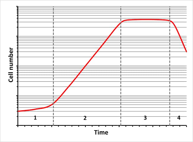
Cell cultures are used in biotechnology to cultivate large numbers of cells under controlled conditions. These cells are then used in the R&D area or for the manufacture of e.g. biotech products, vaccines, or antibodies. The proliferation of these cells takes place over several phases (lag phase, log phase or exponential (growth) phase, stationary phase, death phase) and can be influenced by the targeted supply of nutrients. Many applications make use of the determination of the cell density or cell number to obtain important information on the current status of a cell culture or bacterial suspension. In this method, the measurement is repeated at regular temporal and the results are used to plot a growth curve.1

Figure 1.Bacterial cell growth curve (1 = lag phase; 2 = log phase; 3 = stationary phase; 4 = death phase)
A simple method for determining the cell density is the spectrophotometric measurement of the absorption at a wavelength of 600 nm, known in scientific circles as the “OD600” measurement. In this spectrophotometric measurement, the attenuation of the light intensity caused by the cells present in the culture medium is determined by measuring the absorption.
The attenuation of the light intensity is primarily due to the scatter of the light and only partly to the absorption of the light by the cells itself. This is why the term “optical density” is also used in this context. The optical density shows an excellent correlation to the cell density of dilute cell suspensions up to an OD600 value ≈ 1. Concentrated suspensions should be diluted prior to measurement.2
It should be noted that the measurement of the optical density according to the light-scattering principle on spectrophotometers from different suppliers or of different types can lead to slightly different results. This is due to type-specific factors such as the light source (lamp), the shape and size of the light beam, the detector, or the spectral bandwidth of the spectrophotometer used.
Experimental
This application note describes the determination of the cell number or cell density of cell cultures and bacterial suspensions on the basis of the OD600 value.
The “OD600” method can be used to yield a simple and swift estimate of the cell number. This method is preprogrammed in the Spectroquant® Prove 300 plus and Prove 600 plus UV/VIS spectrophotometers and in the Spectroquant® Prove 100 plus VIS spectrophotometer with firmware version 1.5 or above. No further reagents are required to run this method.
Method
- Spectrophotometric measurement at 600 nm
Measuring range
- -0.020 – 1.200 OD600
Sample material
- Cell cultures, bacterial suspensions
Reagents, Instruments, and Materials
Instruments
For the protein measurement one of the following Spectroquant® photometers is necessary:
- Spectroquant® UV/VIS Spectrophotometer Prove 600 plus (1.73028)
- Spectroquant® UV/VIS Spectrophotometer Prove 300 plus (1.73027)
- Spectroquant® VIS Spectrophotometer Prove 100 plus (1.73026)
Note: Legacy instruments Prove 100/300/600 are also suitable
Software for data maintenance
The Spectroquant® Prove Connect to LIMS software package provides an easy way to transfer your data into a preexisting LIMS system. This software can be purchased under:
- Prove Connect to LIMS (Y11086)
Materials
- Rectangular cells 10 mm (glass) (1.14946)
Analytical approach
Sample Preparation
- Homogenize samples by swirling carefully.
- Avoid shaking the sample too vigorously, since this may lead to damage to the cells.
- Dilute samples with a high cell density or an OD600 value greater than 1.0 with medium or a suitable solution. Do not use distilled water for dilution, since this may lead to damage to the cells and false measurement results.
Measurement
- Open the method list of the photometer and select method No. 313 “OD600”.
- The photometer automatically prompts a zero adjustment. It is recommended to use the same cell for zero adjustment and for sample measurement or a cell with identical optical properties and an identical absorption (matched pair).
- For the zero adjustment fill a 10 mm rectangular cell with cell-culture medium or the solution used for dilution and insert the cell into the cell compartment. The zero adjustment is executed automatically. Confirm the zero adjustment by tapping <OK>. The zero adjustment is valid for the entire measurement series
- Fill the sample solution into a 10 mm rectangular cell and insert the cell into the cell compartment. The measurement starts automatically, and the result is shown in the display.
Data transfer from Prove spectrophotometers (optional)
After measurement, transfer the values measured on the Prove spectrophotometer using the software “Prove Connect to LIMS”.
Cell density
A wide number of calculation tools can be found on the internet that can be used to determine the OD600 result.4 In many cases, these calculation tools are based on empirically determined correlations between the number of cells in E. coli cell cultures and the measured OD600 value. Up to an OD600 value of approx. 1.0, the OD600 value is in excellent correlation with the biomass or the dry mass of the cells. The literature includes many indications that the optical system of the spectrophotometer used also has an impact on the measurement of the OD600 value based on the scatter of light3. This means that while the OD600 method enables a rough estimate of the cell number, the dry mass of the cells – which is dependent on the form and size of the cells – and the optical system of the spectrophotometer must also be taken into account for a more exact determination. This can be achieved e.g. by the parallel microbiological count of the cells in a bacterial culture by plating on culture media and setting the result in relation to the measured OD600 value and determining the corresponding conversion factor.2
Example for an E.coli cell culture:
OD600 = 1.0 corresponds to a cell count of approx. 8 x 10⁸ cells/mL
Conclusion
This method offers a swift and simple alternative for estimating the cell concentration in suspensions. The measurement can be performed without high instrumental expense or additional reagents.
The method is preprogrammed in the Spectroquant® Prove 100 plus, Prove 300 plus, and Prove 600 plus spectrophotometers (also the legacy Prove 100/300/600 instruments).
REFERENCES
如要继续阅读,请登录或创建帐户。
暂无帐户?