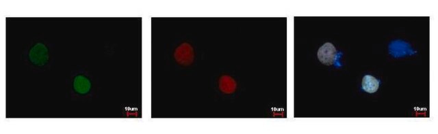
Red fluorescent protein (RFP) is obtained from the Discosoma coral. It can be used as a fluorescent probe for optical microscopy and acts as a biological marker for biomolecule imaging. RFPs exhibit fluorescence emission beyond 57 0nm. They have lower light-scattering, low photo-toxicity and less autofluorescence interference of cells. RFPs are extremely useful for whole body and deep tissue imaging.
Immunogen
Recombinant protein conjugated synthetic peptide encompassing a sequence within the center region of RFP.
Application
Suggested starting dilutions are as follows: IP: 1:100-1:500, WB: 1:5000-1:20000. Not yet tested in other applications. Optimal working dilutions should be determined experimentally by the end user. Monoclonal Anti-RFP antibody produced in mouse has been used in chromatin immunoprecipitation (ChIP) assay.
Features and Benefits
Evaluate our antibodies with complete peace of mind. If the antibody does not perform in your application, we will issue a full credit or replacement antibody. Learn more.
Other Notes
Purification: Affinity purified by Protein G
Physical form
Phosphate-buffered saline, no preservative added.
Disclaimer
Unless otherwise stated in our catalog or other company documentation accompanying the product(s), our products are intended for research use only and are not to be used for any other purpose, which includes but is not limited to, unauthorized commercial uses, in vitro diagnostic uses, ex vivo or in vivo therapeutic uses or any type of consumption or application to humans or animals.
Mitochondrial transport relies on a motor-adaptor complex containing Miro1, a mitochondrial outer membrane protein with two GTPase domains, and TRAK1/2, kinesin-1, and dynein. Using a peroxisome-directed Miro1, we quantified the ability of GTPase mutations to influence the peroxisomal recruitment of
Our team of scientists has experience in all areas of research including Life Science, Material Science, Chemical Synthesis, Chromatography, Analytical and many others.