Skip to Content
Merck
All Photos(4)

Key Documents

H7790

Sigma-Aldrich

Anti-HRD1/SYVN1 antibody produced in rabbit

~1.0 mg/mL, affinity isolated antibody, buffered aqueous solution

Synonym(s):

Anti-Synovial apoptosis inhibitor 1, Anti-Synoviolin, Anti-Synoviolin 1

Sign Into View Organizational & Contract Pricing


About This Item

UNSPSC Code:
12352203
NACRES:
NA.41

biological source

rabbit

conjugate

unconjugated

antibody form

affinity isolated antibody

antibody product type

primary antibodies

clone

polyclonal

form

buffered aqueous solution

mol wt

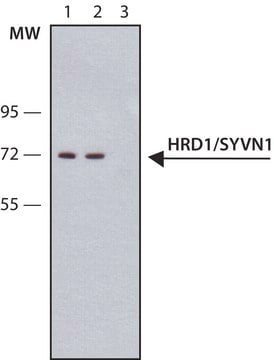
antigen ~70 kDa

species reactivity

mouse, human

concentration

~1.0 mg/mL

technique(s)

immunoprecipitation (IP): 5-10 μg using lysate of mouse brain
indirect immunofluorescence: suitable
western blot: 2-4 μg/mL using whole extract of HEK-293T cells expressing human HRD1/SYVN1.

UniProt accession no.

shipped in

dry ice

storage temp.

−20°C

target post-translational modification

unmodified

Gene Information

human ... SYVN1(84447)
mouse ... Syvn1(74126)
rat ... Syvn1(361712)

General description

Synoviolin (SYVN1) is an endoplasmic reticulum (ER)-membrane resident E3 ubiquitin ligase. The SYVN1 gene is mapped to human chromosome 11q13.1. Also known as HRD1, this protein has five transmembrane domains and a really interesting new gene (RING) finger domain.

Specificity

Anti-HRD1/SYVN1 recognizes human and mouse HRD1/SYVN1.

Application

Anti-HRD1/SYVN1 antibody produced in rabbit has been used in immunoblotting and immunoprecipitation.

Biochem/physiol Actions

Synoviolin (SYVN1) protects against endoplasmic reticulum (ER) stress-induced apoptosis through ER stress-associated protein degradation (ERAD). The really interesting new gene (RING) finger domain of SYVN1 mediates the transfer of ubiquitin from E2 to substrates and a proline-rich domain. E3 ubiquitin-protein ligase (HRD1) expression is strongly induced by ER stress. HRD1 interacts with parkin-associated endothelin receptor-like receptor (Pael-R), promoting its degradation, and protects neurons from cell death caused by the accumulation of Pael-R. HRD1 is also found to enhance the degradation and suppress the toxicity of polyglutamine-expanded huntingtin. In addition to its role in ERAD, HRD1 targets tumor suppressor gene p53 for proteasomal degradation. Overexpression of HRD1 is implicated in the pathogenesis of rheumatoid arthritis. The deletion of gene locus of SYVN1 is implicated in spastic paraplegia, optic atrophy, and neuropathy (SPOAN) disorder.

Physical form

Solution in 0.01 M phosphate buffered saline pH 7.4, containing 15 mM sodium azide.

Storage and Stability

For continuous use, store at 2–8 °C for up to one month. For extended storage, freeze in working aliquots at –20 °C. Repeated freezing and thawing is not recommended. If slight turbidity occurs upon prolonged storage, clarify the solution by centrifugation before use. Working dilution samples should be discarded if not used within 12 hours.

Disclaimer

Unless otherwise stated in our catalog or other company documentation accompanying the product(s), our products are intended for research use only and are not to be used for any other purpose, which includes but is not limited to, unauthorized commercial uses, in vitro diagnostic uses, ex vivo or in vivo therapeutic uses or any type of consumption or application to humans or animals.

Not finding the right product?  

Try our Product Selector Tool.

Storage Class Code

10 - Combustible liquids

Flash Point(F)

Not applicable

Flash Point(C)

Not applicable

Personal Protective Equipment

dust mask type N95 (US), Eyeshields, Gloves

Certificates of Analysis (COA)

Search for Certificates of Analysis (COA) by entering the products Lot/Batch Number. Lot and Batch Numbers can be found on a product’s label following the words ‘Lot’ or ‘Batch’.

Already Own This Product?

Find documentation for the products that you have recently purchased in the Document Library.

Visit the Document Library

Kit Briant et al.
Journal of cell science, 128(22), 4112-4125 (2015-10-09)
Clearance of misfolded proteins from the endoplasmic reticulum (ER) is mediated by the ubiquitin-proteasome system in a process known as ER-associated degradation (ERAD). The mechanisms through which proteins containing aberrant transmembrane domains are degraded by ERAD are poorly understood. To
Tomohiro Omura et al.
Journal of neuroscience research, 86(7), 1577-1587 (2008-02-05)
HRD1 is an E3 ubiquitin ligase and plays an important role in endoplasmic reticulum-associated degradation (ERAD). Parkin-associated endothelin receptor-like receptor (Pael-R) is a substrate of the E3 ubiquitin ligase parkin, which has been implicated in ER stress-induced cell death in
Jeffrey M Lynch et al.
Cell, 149(6), 1257-1268 (2012-06-12)
Thrombospondin (Thbs) proteins are induced in sites of tissue damage or active remodeling. The endoplasmic reticulum (ER) stress response is also prominently induced with disease where it regulates protein production and resolution of misfolded proteins. Here we describe a function
Hui Yang et al.
Experimental cell research, 313(3), 538-550 (2006-12-05)
E3 ubiquitin ligases catalyze the conjugation of ubiquitin onto proteins, which acts as a signal for targeting proteins for degradation by the proteasome. Hrd1 is an endoplasmic reticulum (ER) membrane-spanning E3 with its catalytic active RING finger facing the cytosol.
Yupeng Zhao et al.
iScience, 23(9), 101494-101494 (2020-09-12)
Atlastin (ATL) is a class of dynamin-like GTPases shaping endoplasmic reticulum (ER) by mediating homotypic membrane fusion. Defect of ATLs leads to abnormal ER structure and hereditary spastic paraplegia (HSP), a neurodegenerative disease with progressive spasticity. How ATLs are regulated

Our team of scientists has experience in all areas of research including Life Science, Material Science, Chemical Synthesis, Chromatography, Analytical and many others.

Contact Technical Service