Skip to Content
Merck
All Photos(7)

Documents

C9848

Sigma-Aldrich

Monoclonal Anti-Calbindin-D-28K antibody produced in mouse

enhanced validation

clone CB-955, ascites fluid

Synonym(s):

Calbindin Antibody, Calbindin Antibody - Monoclonal Anti-Calbindin-D-28K antibody produced in mouse

Sign Into View Organizational & Contract Pricing


About This Item

MDL number:
UNSPSC Code:
12352203
NACRES:
NA.44

biological source

mouse

Quality Level

conjugate

unconjugated

antibody form

ascites fluid

antibody product type

primary antibodies

clone

CB-955, monoclonal

mol wt

antigen 28 kDa

contains

15 mM sodium azide

species reactivity

mouse, human, guinea pig, canine, sheep, rat, pig, rabbit, feline, bovine, goat

enhanced validation

independent
Learn more about Antibody Enhanced Validation

concentration

15-55 mg/mL

technique(s)

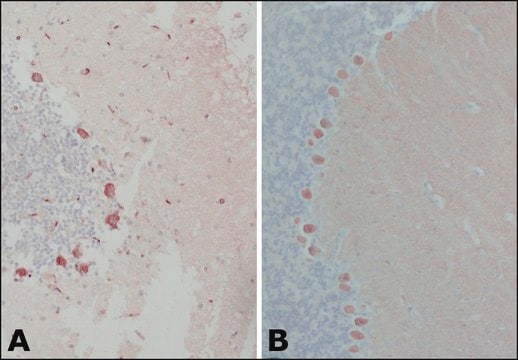
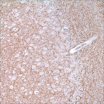
immunohistochemistry (formalin-fixed, paraffin-embedded sections): 1:3,000 using rat cerebellum sections
indirect ELISA: suitable
western blot: 1:3,000 using MDBK cell extract

isotype

IgG1

UniProt accession no.

shipped in

dry ice

storage temp.

−20°C

target post-translational modification

unmodified

Gene Information

human ... CALB1(793)
mouse ... Calb1(12307)
rat ... Calb1(83839)

General description

Calbindin-D-28K is a highly conserved 28 kDa calcium binding protein with broad tissue distribution.

Monoclonal Anti-Calbindin-D-28K (mouse IgG1 isotype) is derived from the CB-955 hybridoma produced by the fusion of mouse myeloma cells and splenocytes from BALB/c mice immunized with a purified bovine kidney calbindin-D-28K. The isotype is determined by a double diffusion immunoassay using Mouse Monoclonal Antibody Isotyping Reagents, Catalog Number ISO2.

Calbindin-D-28K belongs, together with calmodulin, S-100, parvalbumin, troponin C and other proteins, to a family of low molecular weight calcium-binding proteins (CaBPs). These CaBPs have homologous primary structures, which contain polypeptide folds of the EF-hand type for the acceptance of Ca2+. Calbindin is an attractive neuroanatomical marker. Despite its broad tissue distribution, calbindin-D-28K exhibits a cell-type specific expression pattern. Thus, it has been immunocytochemically localized in selected cells in many CNS structures, where it is thought to act either as an intracellular calcium buffer, or in the intramembranous transport of calcium. It is found predominantly in subpopulations of central and peripheral nervous system neurons, and in certain epithelial cells involved in Ca2+ transport such as distal tubular cells and cortical collecting tubules of the kidney, and in enteric neuroendocrine cells. Monoclonal antibody reacting specifically with calbindin-D-28K is instrumental in the localization of calbindin in different tissues and cell populations.

Specificity

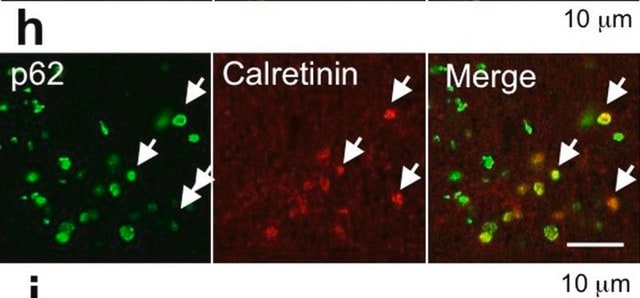
The antibody does not react with other members of the EF-hand family, such as calbindin-D-9K, calretinin, myosin light chain, parvalbumin, S-100a, S-100b, S-100A2 (S100L) and S-100A6 (calcyclin). A weaker reactivity was observed with chicken calbindin-D-28K.

Immunogen

Bovine kidney calbindin-D

Application

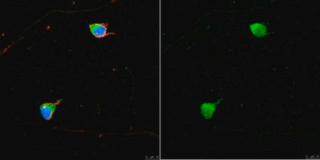
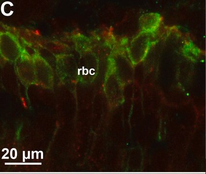
Monoclonal Anti-Calbindin-D-28K antibody produced in mouse may be used in immunohistochemistry (alcohol-, formalin-, and methacarn-fixed, paraffin-embedded, and frozen tissue sections). Enzymatic predigestion of formalin-fixed, paraffin-embedded sections improves immunohistochemical staining. The antibody produced in mouse is suitable for indirect ELISA, western blotting at a working dilution of 1:3000 using MDBK cell extract and indirect immunoperoxidase staining with a working dilution of 1:3000 using trypsin-digested, formalin-fixed, paraffin-embedded rat cerebellum sections. It was used for immunohistochemistry and immunocytochemistry at a working dilution of 1:500 in a study to understand impaired cerebellar development and function in mice lacking CAPS2, a protein involved in neurotrophin release. It was used for immunostaining to gauge the development of neuronal and glial cells in the inner nuclear layer of islet-class factor Islet-1 (Isl1)-null and control retinas. It was used for immunohistochemical colocalization studies to confirm postsynaptic localization of the RGS11 complex in a study.

Biochem/physiol Actions

Calbindin-D-28K is involved in intramembranous transport of calcium or acts as an intracellualr calcium buffer in many CNS structures such as subpopulations of central and peripheral nervous system neurons. It is also present in certain epithelial cells involved in Ca2+ transport such as distal tubular cells and cortical collecting tubules of the kidney, and in enteric neuroendocrine cells.

Physical form

Reagent is supplied as ascites fluid with 15mM sodium azide as a preservative.

Storage and Stability

For continuous use, store at 2-8 °C for up to one month. For extended storage, freeze in working aliquots. Repeated freezing and thawing, or storage in "frost free" freezers, is not recommended. If slight turbidity occurs upon prolonged storage, clarify the solution by centrifugation before use. Working dilution samples should be discarded if not used within 12 hours.

Disclaimer

Unless otherwise stated in our catalog or other company documentation accompanying the product(s), our products are intended for research use only and are not to be used for any other purpose, which includes but is not limited to, unauthorized commercial uses, in vitro diagnostic uses, ex vivo or in vivo therapeutic uses or any type of consumption or application to humans or animals.

Not finding the right product?  

Try our Product Selector Tool.

Storage Class Code

10 - Combustible liquids

WGK

WGK 1


Certificates of Analysis (COA)

Search for Certificates of Analysis (COA) by entering the products Lot/Batch Number. Lot and Batch Numbers can be found on a product’s label following the words ‘Lot’ or ‘Batch’.

Already Own This Product?

Find documentation for the products that you have recently purchased in the Document Library.

Visit the Document Library

T G Ohm et al.
Neuroscience, 42(3), 823-840 (1991-01-01)
Calbindin-D-28k and parvalbumin are calcium-binding proteins. The laminar distribution and morphological features of calbindin-D-28k-like immunoreactive structures were studied in 60-microns-thick sections of the human olfactory bulb. Except for the olfactory nerve layer, immunoreactive neurons were present in all layers of
Bruno Cozzi et al.
Frontiers in systems neuroscience, 8, 42-42 (2014-04-16)
The mammalian claustrum is involved in processing sensory information from the environment. The claustrum is reciprocally connected to the visual cortex and these projections, at least in carnivores, display a clear retinotopic distribution. The visual cortex of dolphins occupies a
L M Garcia-Segura et al.
Brain research, 296(1), 75-86 (1984-03-26)
A complete mapping of immunoreactive sites for vitamin D-dependent calcium-binding protein (CaBP) was performed on serial sections from the rat central nervous system. CaBP immunoreactivity was found in the perikarya, dendrites and axons of some neurons from the limbic system
Yasser Elshatory et al.
The Journal of neuroscience : the official journal of the Society for Neuroscience, 27(46), 12707-12720 (2007-11-16)
Whereas the mammalian retina possesses a repertoire of factors known to establish general retinal cell types, these factors alone cannot explain the vast diversity of neuronal subtypes. In other CNS regions, the differentiation of diverse neuronal pools is governed by
Larisa C Kruger et al.
The Journal of neuroscience : the official journal of the Society for Neuroscience, 36(23), 6213-6224 (2016-06-10)
Voltage-gated sodium channel (VGSC) β subunits signal through multiple pathways on multiple time scales. In addition to modulating sodium and potassium currents, β subunits play nonconducting roles as cell adhesion molecules, which allow them to function in cell-cell communication, neuronal

Our team of scientists has experience in all areas of research including Life Science, Material Science, Chemical Synthesis, Chromatography, Analytical and many others.

Contact Technical Service