DUO94004
Duolink® flowPLA Detection Kit - FarRed
Duolink® PLA kit for Flow Cytometry with FarRed Detection
Synonym(s):
in situ Proximity Ligation Assay, Flowcytometry-PLA, Protein Protein Interaction Kit
Sign Into View Organizational & Contract Pricing
All Photos(4)
About This Item
UNSPSC Code:
41105331
NACRES:
NA.32
Recommended Products
product line
Duolink®
technique(s)
flow cytometry: suitable
immunofluorescence: suitable
proximity ligation assay: suitable
fluorescence
λex 644 nm; λem 669 nm
suitability
suitable for fluorescence
shipped in
dry ice
storage temp.
−20°C
General description
Duolink® flowPLA Detection Kit contains detection oligonucleotides with a fluorophore (lex = 644 nm/lem = 669 nm).
Specificity
Far Red Fluorescence Detection Reagents
Use appropriate laser for λex 644 nm excitation
Use appropriate filter for λem 669 nm emission
Use appropriate laser for λex 644 nm excitation
Use appropriate filter for λem 669 nm emission
Application
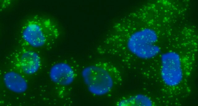
Based on proximity ligation assay (PLA), the Duolink® PLA Technology allows for endogenous detection of protein interactions, post-translational modifications, and protein expression levels at the single molecule level in fixed cells.
Duolink® flowPLA Detection Kits will enable sensitive detection of proteins, protein-protein interactions, and protein modifications within cell populations by flow cytometry. To perform a Duolink® flowPLA experiment, you will need fixed, suspended cells, two primary antibodies that specifically recognize your proteins of interest, a pair of PLA probes (one 100RXN PLUS and one 100RXN MINUS), wash buffer, and a Duolink® flowPLA Detection Kit. The flowPLA Kits are available with 5 different fluorophores: Violet, Red, Green, Orange, or FarRed. The flowPLA Kits contain all the necessary reagents to perform the amplification and detection of bound PLA probes by flow cytometry. Analysis is carried out using standard flow cytometry assay equipment. User must provide a fixed cell suspension, primary antibodies, and corresponding PLA Probes.
Follow the Duolink® PLA Flow Cytometry Protocol to use this product.
Visit our Duolink® PLA Flow Cytometry page on how to run a Duolink® flow experiment, applications, troubleshooting, and more.
Application Note
Primary antibodies are needed. Test your primary antibodies (IgG-class, mono- or polyclonal) in a standard immunofluorescence (IF), immunohistochemistry (IHC), or immunocytochemistry (ICC) assay to determine the optimal fixation, blocking, and titer conditions. Flow validated antibodies are recommended.
Let us do the work for you, learn more about our Custom Service Program to accelerate your Duolink® projects
View full Duolink® product list
Duolink® flowPLA Detection Kit–FarRed has been used in in situ proximity ligation assay:
- to detect interaction and complex formation between cellular retinoic acid binding protein 1 (Crabp1) and the components of rapidly accelerated fibrosarcoma (Raf) kinase- MAPK-Erk kinase (Mek) signaling pathway
- to study protein interaction in human cultured MOLT-4 cells and HeLa cells
- to visualize Beclin-1 protein interaction with 14-3-3t in neurons
- to study protein interactions in graft endothelial cells
Features and Benefits
- Analyze protein protein interactions with flow cytometry readout
- Analyze cell populations with Proximity Ligation Assay
- Increased sensitivity due to rolling circle amplification for low abundant targets
- No overexpression or genetic manipulation required
- Relative quantification possible
- Works with any flow cytometer instrumentation
- Easy to follow flexible protocol
- Publication-ready results
Components
This product is comprised of the following:
See datasheet for more information.
- 5x Detection Solution - FarRed (DUO84004)
- 5x Ligation Buffer (DUO82009)
- 5x Amplification Buffer (DUO82050)
- Ligase (1U/μL)
- Polymerase (10U/μL)
See datasheet for more information.
Legal Information
Duolink is a registered trademark of Merck KGaA, Darmstadt, Germany
PLA is a registered trademark of Merck KGaA, Darmstadt, Germany
Signal Word
Danger
Hazard Statements
Precautionary Statements
Hazard Classifications
Resp. Sens. 1
Storage Class Code
10 - Combustible liquids
Certificates of Analysis (COA)
Search for Certificates of Analysis (COA) by entering the products Lot/Batch Number. Lot and Batch Numbers can be found on a product’s label following the words ‘Lot’ or ‘Batch’.
Already Own This Product?
Find documentation for the products that you have recently purchased in the Document Library.
Customers Also Viewed
Adriana Cassaro et al.
Hematological oncology, 39(3), 364-379 (2021-01-27)
Wnt/Fzd signaling has been implicated in hematopoietic stem cell maintenance and in acute leukemia establishment. In our previous work, we described a recurrent rearrangement involving the WNT10B locus (WNT10BR ), characterized by the expression of WNT10BIVS1 transcript variant, in acute
Tyler J Burns et al.
Cytometry. Part A : the journal of the International Society for Analytical Cytology, 91(2), 180-189 (2017-01-18)
To quantify visual and spatial information in single cells with a throughput of thousands of cells per second, we developed Subcellular Localization Assay (SLA). This adaptation of Proximity Ligation Assay expands the capabilities of flow cytometry to include data relating
Sofie Selmer Andersen et al.
Cytokine, 64(1), 54-57 (2013-06-04)
Many cytokine receptors are cell surface proteins that promiscuously combine to form active signalling homo- or heterodimers. Thus, receptor chain dimerization can be viewed as a direct measure of a high probability of intracellular signalling by specific cytokines. Proximity ligation
Sung Wook Park et al.
Scientific reports, 9(1), 10929-10929 (2019-07-31)
The rapidly accelerated fibrosarcoma (Raf) kinase is canonically activated by growth factors that regulate multiple cellular processes. In this kinase cascade Raf activation ultimately results in extracellular regulated kinase 1/2 (Erk1/2) activation, which requires Ras binding to the Ras binding
Karl-Johan Leuchowius et al.
Cytometry. Part A : the journal of the International Society for Analytical Cytology, 75(10), 833-839 (2009-08-04)
Interactions between members of the epidermal growth factor receptor (EGFR) family mediates cellular responses to ligand stimulation. Measurement of these interactions could provide important information and may prove useful as prognostic markers in malignancy. Therefore, to develop methods to study
Our team of scientists has experience in all areas of research including Life Science, Material Science, Chemical Synthesis, Chromatography, Analytical and many others.
Contact Technical Service


