Skip to Content
Merck
All Photos(2)

Key Documents

MABN71

Sigma-Aldrich

Anti-GluR2 Antibody, clone L21/32

clone L21/32, from mouse

Synonym(s):

glutamate receptor, ionotropic, AMPA 2, glutamate receptor 2, Glutamate receptor ionotropic, AMPA 2, AMPA-selective glutamate receptor 2

Sign Into View Organizational & Contract Pricing


About This Item

UNSPSC Code:
12352203
eCl@ss:
32160702
NACRES:
NA.41

biological source

mouse

Quality Level

antibody form

purified antibody

antibody product type

primary antibodies

clone

L21/32, monoclonal

species reactivity

rat

species reactivity (predicted by homology)

mouse (based on 100% sequence homology), human (immunogen homology)

technique(s)

immunohistochemistry: suitable
western blot: suitable

isotype

IgG1κ

NCBI accession no.

UniProt accession no.

shipped in

wet ice

target post-translational modification

unmodified

Gene Information

human ... GRIA2(2891)

General description

Glutamate receptors (GluRs) can be categorized as ionotropic or metabotropic and subcatergorized by their agonist preferences (NMDA, AMPA or Kainic acid). There are four types of AMPA selective GluR subunits (GluR1, GluR2, GluR3 and GluR4). Tetrameric or pentameric combinations of different subunits contributes to the functional diversity of AMPA receptors. In general, AMPA receptors mediate fast synaptic current at most excitatory synapses, with stoichiometry characterized by subtype composition. Although subunit composition of AMPA receptors varies, they must contain at least one edited GluR2 subunit to be calcium impermeable. The critical residue controlling calcium permeability is in the pore loop region. In GluR1, GluR3, and GluR4, this position is occupied by a Gln residue. In GluR2, it is occupied by an Arg residue. It has been shown experimentally that the presence of Arg in this position blocks Ca2+ ion permeability, while a Gln does not. Relative calcium permeability in AMPA receptor channels may be significant in pathological neurotoxic damage and long term changes in nervous system responses.

Immunogen

Recombinant protein corresponding to human GluR2.

Application

Detect GluR2 using this Anti-GluR2 Antibody, clone L21/32 validated for use in WB, IH.
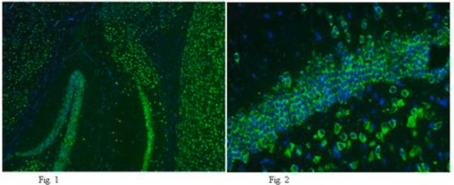
Immunohistochemistry Analysis: 1:300 dilution of this antibody from a previous lot detected GluR2 in rat cerebellum and hippocampus tissue.
Research Category
Neuroscience

Neuroscience
Research Sub Category
Neurodegenerative Diseases

Neurotransmitters & Receptors

Quality

Evaluated by Western Blot in rat brain membrane tissue lysate.

Western Blot Analysis: 0.5 µg/mL of this antibody detected GluR2 on 10 µg of rat brain membrane tissue lysate.

Target description

~ 96 kDa observed

Physical form

Format: Purified
Protein G
Purified mouse monoclonal IgG1κ in buffer containing 0.1 M Tris-Glycine (pH 7.4), 150 mM NaCl with 0.05% sodium azide.

Storage and Stability

Stable for 1 year at 2-8°C from date of receipt.

Analysis Note

Control
Rat brain membrane tissue lysate

Other Notes

Concentration: Please refer to the Certificate of Analysis for the lot-specific concentration.

Disclaimer

Unless otherwise stated in our catalog or other company documentation accompanying the product(s), our products are intended for research use only and are not to be used for any other purpose, which includes but is not limited to, unauthorized commercial uses, in vitro diagnostic uses, ex vivo or in vivo therapeutic uses or any type of consumption or application to humans or animals.

Not finding the right product?  

Try our Product Selector Tool.

Storage Class Code

12 - Non Combustible Liquids

WGK

WGK 1

Flash Point(F)

Not applicable

Flash Point(C)

Not applicable


Certificates of Analysis (COA)

Search for Certificates of Analysis (COA) by entering the products Lot/Batch Number. Lot and Batch Numbers can be found on a product’s label following the words ‘Lot’ or ‘Batch’.

Already Own This Product?

Find documentation for the products that you have recently purchased in the Document Library.

Visit the Document Library

Venkat Swaroop Achuta et al.
Science signaling, 11(513) (2018-01-18)
Altered neuronal network formation and function involving dysregulated excitatory and inhibitory circuits are associated with fragile X syndrome (FXS). We examined functional maturation of the excitatory transmission system in FXS by investigating the response of FXS patient-derived neural progenitor cells
Maria Arvaniti et al.
Journal of neurochemistry, 138(6), 806-820 (2016-06-28)
Nicotinic acetylcholine receptors (nAChRs) affect multiple physiological functions in the brain and their functions are modulated by regulatory proteins of the Lynx family. Here, we report for the first time a direct interaction of the Lynx protein LY6/PLAUR domain-containing 6
Joseph A McQuail et al.
Neuropharmacology, 197, 108720-108720 (2021-07-18)
Ionotropic glutamate receptors of the NMDA and AMPA subtypes transduce excitatory signaling on neurons in the prefrontal cortex (PFC) in support of cognitive flexibility. Cognitive flexibility is reliably observed to decline at advanced ages, coinciding with changes in PFC glutamate
Identification of a Glutamatergic Claustrum-Anterior Cingulate Cortex Circuit for Visceral Pain Processing.
Xu, et al.
The Journal of Neuroscience, 42, 8154-8168 (2023)
Xiang-Dong Sun et al.
Nature neuroscience, 19(8), 1010-1018 (2016-06-14)
Neurotransmission requires precise control of neurotransmitter release from axon terminals. This process is regulated by glial cells; however, the underlying mechanisms are not fully understood. We found that glutamate release in the brain was impaired in mice lacking low-density lipoprotein

Our team of scientists has experience in all areas of research including Life Science, Material Science, Chemical Synthesis, Chromatography, Analytical and many others.

Contact Technical Service