Flow Cytometry Guide
Flow cytometry is a laboratory technique that is used to analyze cells. Reviewing the basics of flow cytometry, immunodetection methods, key flow cytometry protocol steps, and proper controls helps to ensure accurate flow cytometric analysis. Read on to uncover tips that will make your next flow cytometry experiment successful.
What Is Flow Cytometry?
Flow cytometry is a powerful technique for characterizing and/or sorting heterogeneous suspended cell populations based on their physical and fluorescence characteristics. It can be considered a form of single-cell analysis as flow cytometry is well suited for experiments where quantitative information is desired at a single-cell level for large amounts of cells simultaneously. This can provide information on the number of cells of a given type and the amount of a specific protein in the cell.
Some benefits of flow cytometry include quick analysis, calling attention to differences in cell subpopulations when looking at heterogeneous cell populations, and analyzing subpopulations of heterogeneous cell populations. Flow cytometry is commonly used in immunophenotyping and immuno-oncology research applications to analyze hematological differences in blood, bone marrow, and other sample types.
How It Works
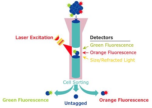
The flow cytometry process works by relying on hydrodynamic focusing of a cell suspension sample to create a single-cell stream, which passes in front of a laser. How the cell scatters incident light is used to determine the size and intracellular complexity of cells at a rate of thousands of particles per second. The analog data generated is then converted to digital data that can be quantified and plotted.
From flow cytometric measurements, specific populations of cells and subsets within these cells can be defined and physically isolated via cell sorting, where cell charge is manipulated based on fluorescence characteristics to allow electrostatic deflection of particles. More precise identification of subpopulations requires the use of probes. Adherent cells and solid tissues may also be analyzed by this technique if they can be successfully dissociated to create a single-cell suspension.

Figure 1.Diagram of typical flow cytometric analysis.
Components of a Traditional Flow Cytometer
For flow cytometric analysis, at a minimum, even basic, single-laser flow cytometers are typically equipped with sufficient filters to permit the detection of four fluorophores simultaneously. And, it is currently possible to differentiate as many as 18 wavelengths of light in a single experiment. The components of a traditional flow cytometer include:
Fluidics for Sample Processing and Cell Sorting
- Flow Chamber: Cells in the sample are hydrodynamically focused and presented to the laser.
Optics for Fluorescence Detection
- Light moves through a system of mirrors and filters.
- Cells can be measured based on their size, internal complexity, and signal intensity.
Electronics for Signal Processing, Computer Display, and Analysis
- Photomultiplier tube converts light into electrical signals.
- Signal is amplified and digitized.
Immunodetection by Flow Cytometry
As with other immunodetection applications, there are several approaches to the use of antibodies to probe for specific cell moieties by flow cytometry. Two main methods are commonly employed: direct detection and indirect detection.
Direct Detection
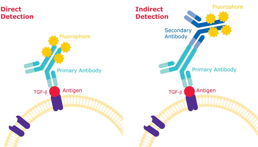
Direct detection refers to a single-step staining process, which employs a primary antibody that specifically binds to an epitope of interest and that is directly conjugated to a molecule that permits visualization or other detection of the binding event. When probing for antigens localized to the cell surface, fixation of cells is not recommended, as this process renders antigens of interest inaccessible to antibody probes. Hence, it is important to keep unfixed cells viable until the data acquisition is complete. To find antibodies for direct detection, search our Antibody Explorer tool by setting the Search Within to “Primary Antibodies Only” and selecting the application “flow cytometry".
Indirect Detection
In the indirect detection method, incubation with a purified antibody to permit binding to an antigen of interest is followed by a fluorophore-conjugated secondary antibody specific for the primary antibody host isotype, forming a primary-fluorescent secondary antibody scaffold. Increased modularity in an antibody library may be achieved by the use of purified primary antibodies and fluorophore-conjugated secondary antibodies in a variety of wavelengths (or colors) and specific for host isotypes in which the primaries are raised. To find antibodies for indirect detection, search the product table on our Secondary Antibodies page and filter the technique to “flow cytometry”.
Learn more about the detection of fluorochromes in flow cytometry with our guides on selecting the right fluorochrome for your flow cytometry experiment and building the optimal panel for multicolor analysis.

Figure 2.Direct detection vs indirect detection methods for flow cytometry.
Key Flow Cytometry Protocol Steps
The four main steps in flow cytometry protocols are:
- Sample Preparation: Single-cell suspensions should be prepped through the use of mechanical dissociation methods and detachment techniques such as using enzymatic solutions or calcium chelation reagents.
- Blocking: Anti-Fc antibody dilution is usually applied to suspended cells to prevent nonspecific binding of primary antibody.
- Antibody Incubation: Flow cytometry incubation can involve various components such as primary antibodies, secondary antibodies, streptavidin, and fluorochromes.
- Data Acquisition: Most flow cytometers are accompanied by the necessary software to obtain and transform the signals from cell characteristics. The user then sets the specific software parameters for their experiment.
Discover more details on these steps in our Key Steps in Flow Cytometry Protocol article, or explore troubleshooting tips in our
Flow Cytometry Troubleshooting Guide.
Proper Controls
In addition to cells of interest, every flow cytometry experiment should include the following controls:
- At least one unstained sample that has been incubated with buffer at each step at the same time as the test samples to optimize flow rates and voltages in the experiment.
- An appropriate negative control sample, which will be identical to the test samples except for the substitution of the primary antibody with an isotype control that is raised in the same host species as the primary antibody. This controls for nonspecific binding of the secondary antibody. To find isotype controls, browse the table on our Flow Cytometry Antibodies page. You can search the table by keyword, such as “control”.
- Where possible, a positive control that is comprised of cells known to express each antigen of interest, co-incubated with the test samples, and tested with only a single color.
Para seguir leyendo, inicie sesión o cree una cuenta.
¿No tiene una cuenta?