MAB3839-I
Anti-Transglutaminase-2 Antibody, FN binding domain Antibody, clone 4G3
clone 4G3, from mouse
Synonym(s):
Protein-glutamine gamma-glutamyltransferase 2, EC: 2.3.2.13, Tissue transglutaminase, Transglutaminase C, TG(C), TGC, TGase C, Transglutaminase H, TGase H, TGase-2
About This Item
Recommended Products
biological source
mouse
antibody form
purified immunoglobulin
antibody product type
primary antibodies
clone
4G3, monoclonal
species reactivity
human
packaging
antibody small pack of 25 μg
technique(s)
flow cytometry: suitable
immunofluorescence: suitable
immunoprecipitation (IP): suitable
western blot: suitable
isotype
IgG1κ
NCBI accession no.
UniProt accession no.
target post-translational modification
unmodified
Gene Information
human ... TGM2(7052)
General description
Specificity
Immunogen
Application
Signaling
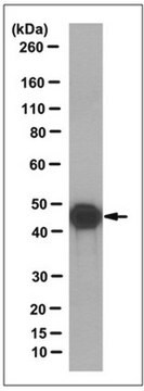
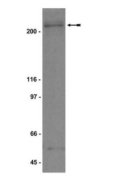
Western Blotting Analysis: A representative lot detected Transglutaminase-2, FN binding domain in Western Blotting applications (Janiak, A., et. al. (2006). Mol Biol Cell. 17(4):1606-19).
Radioimmunoassay Analysis: A representative lot detected Transglutaminase-2, FN binding domain in Radioimmunoassay applications (Akimov, S.S., et. al. (2001). Blood. 98(5):1567-76).
Flow Cytometry Analysis: A representative lot detected Transglutaminase-2, FN binding domain in Flow Cytometry applications (Zemskov, E.A., et. al. (2011). PLos One. 6(4):e19414).
Immunoprecipitation Analysis: A representative lot immunoprecipitated Transglutaminase-2, FN binding domain in Immunoprecipitation applications (Zemskov, E.A., et. al. (2011). PLos One. 6(4):e19414; Janiak, A., et. al. (2006). Mol Biol Cell. 17(4):1606-19).
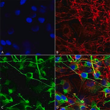
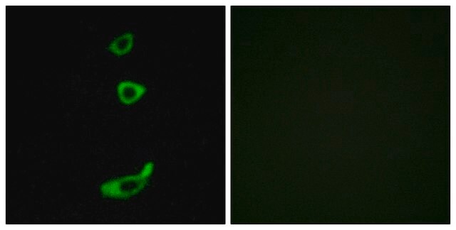
Immunofluorescence Analysis: A representative lot detected Transglutaminase-2, FN binding domain in Immunofluorescence applications (Janiak, A., et. al. (2006). Mol Biol Cell. 17(4):1606-19; Akimov, S.S., et. al. (2001). Blood. 98(5):1567-76; Zemskov, E.A., et. al. (2011). PLos One. 6(4):e19414).
Quality
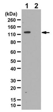
Western Blotting Analysis: 1 µg/mL of this antibody detected Transglutaminase-2, FN binding domain in A431 cell lysate.
Target description
Linkage
Physical form
Storage and Stability
Handling Recommendations: Upon receipt and prior to removing the cap, centrifuge the vial and gently mix the solution. Aliquot into microcentrifuge tubes and store at -20°C. Avoid repeated freeze/thaw cycles, which may damage IgG and affect product performance.
Other Notes
Disclaimer
Not finding the right product?
Try our Product Selector Tool.
Certificates of Analysis (COA)
Search for Certificates of Analysis (COA) by entering the products Lot/Batch Number. Lot and Batch Numbers can be found on a product’s label following the words ‘Lot’ or ‘Batch’.
Already Own This Product?
Find documentation for the products that you have recently purchased in the Document Library.
Our team of scientists has experience in all areas of research including Life Science, Material Science, Chemical Synthesis, Chromatography, Analytical and many others.
Contact Technical Service