Skip to Content
Merck
All Photos(3)

Documents

16-232

Sigma-Aldrich

Anti-α-Tubulin Antibody, clone DM1A, Alexa Fluor 488 conjugate

clone DM1A, Upstate®, from mouse

Synonym(s):

Alpha-tubulin 3, Tubulin B-alpha-1, Tubulin alpha-3 chain, tubulin, alpha 1a, tubulin, alpha 3, tubulin, alpha, brain-specific

Sign Into View Organizational & Contract Pricing


About This Item

UNSPSC Code:
12352203
eCl@ss:
32160702
NACRES:
NA.41

biological source

mouse

Quality Level

conjugate

ALEXA FLUOR 488

antibody form

purified antibody

antibody product type

primary antibodies

clone

DM1A, monoclonal

species reactivity

rat, mouse, guinea pig, human, pig, bovine, avian

manufacturer/tradename

Upstate®

technique(s)

flow cytometry: suitable
immunocytochemistry: suitable
immunofluorescence: suitable
western blot: suitable

isotype

IgG1

NCBI accession no.

UniProt accession no.

shipped in

wet ice

target post-translational modification

unmodified

Gene Information

human ... TUBA1A(7846)

General description

Tubulins, major components of the cellular cytoskeleton, form microtubules that are critical for cellular structure, migration, cell division and intracellular trafficking. There are at least three types of tubulins, alpha, beta and gamma. Each has a molecular weight of approximately 55 kDa. Microtubules are large structures (300 angstrom diameter) composed of alternating alpha and beta subunits. Compounds that block microtuble formation, such as Taxol and the vinca alkaloids, inhibit cell division and are effective anti-cancer agents.

Specificity

Recognizes α-Tubulin, clone DM1A, Mr 50-60 kDa.

Immunogen

Epitope: The epitopehas been mapped to amino acids 426-450 (AALEKDYEEVGVDSV-EGEGEEEGEE).
Purified human brain α-Tubulin. The epitope recognized by this antibody has been mapped to amino acids 426-450 (AALEKDYEEVGVDSV-EGEGEEEGEE).

Application

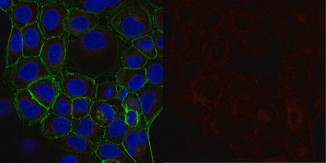
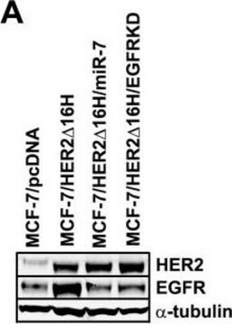
Anti-α-Tubulin Antibody, clone DM1A, Alexa Fluor 488 conjugate is an antibody against α-Tubulin for use in FC, IC, IF & WB.
Western Blot Analysis:
2 µg/mL of a previous lot detected α-Tubulin in RIPA lysates from A431 cells.

Immunocytochemistry:
2 µg/mL of a previous lot showed positive immunostaining for α-Tubulin in HeLa cells.

Quality

Routinely evaluated by Flow Cytometry.

Flow Cytometry:
0.2 μg of this lot detected α-Tubulin in fixed and permeabilized rat L6, Jurkat, and A431 cells.

Target description

50-60 kDa

Physical form

Alexa Fluor 488 conjugated, purified mouse monoclonal IgG1 in buffer containing PBS containing 1% BSA, 0.05% Tween-20, 0.05% sodium azide.
Liquid at 2-8°C.

Storage and Stability

Stable for 1 year at 2-8°C from date of receipt.
Do Not Freeze. Do not store the material diluted.
For maximum recovery of product, centrifuge original vial prior to removing cap.

Analysis Note

Control
Negative Control: Catalog # 16-240, Normal Mouse IgG, Alexa Fluor 488-conjugate.

Other Notes

Concentration: Please refer to the Certificate of Analysis for the lot-specific concentration.

Legal Information

ALEXA FLUOR is a trademark of Life Technologies
UPSTATE is a registered trademark of Merck KGaA, Darmstadt, Germany

Not finding the right product?  

Try our Product Selector Tool.

Storage Class Code

12 - Non Combustible Liquids

WGK

WGK 2

Flash Point(F)

Not applicable

Flash Point(C)

Not applicable


Certificates of Analysis (COA)

Search for Certificates of Analysis (COA) by entering the products Lot/Batch Number. Lot and Batch Numbers can be found on a product’s label following the words ‘Lot’ or ‘Batch’.

Already Own This Product?

Find documentation for the products that you have recently purchased in the Document Library.

Visit the Document Library

Simon Schneider et al.
eLife, 12 (2023-11-28)
Cylicins are testis-specific proteins, which are exclusively expressed during spermiogenesis. In mice and humans, two Cylicins, the gonosomal X-linked Cylicin 1 (Cylc1/CYLC1) and the autosomal Cylicin 2 (Cylc2/CYLC2) genes, have been identified. Cylicins are cytoskeletal proteins with an overall positive
Naila Umer et al.
Frontiers in cell and developmental biology, 9, 749559-749559 (2021-12-07)
Profilins (PFNs) are key regulatory proteins for the actin polymerization in cells and are encoded in mouse and humans by four Pfn genes. PFNs are involved in cell mobility, cell growth, neurogenesis, and metastasis of tumor cells. The testes-specific PFN3
Yin-Wei Kuo et al.
Proceedings of the National Academy of Sciences of the United States of America, 116(12), 5533-5541 (2019-03-07)
The remodeling of the microtubule cytoskeleton underlies dynamic cellular processes, such as mitosis, ciliogenesis, and neuronal morphogenesis. An important class of microtubule remodelers comprises the severases-spastin, katanin, and fidgetin-which cut microtubules into shorter fragments. While severing activity might be expected
cAMP, tubulin, axonal transport, and regeneration.
Liu, Howard H and Brady, Scott T
Experimental neurology, 189, 199-203 (2004)
Atsuko Kubo et al.
The Journal of comparative neurology, 527(5), 985-998 (2018-11-09)
Tau is a microtubule-associated protein (MAP) that is localized to the axon. In Alzheimer's disease (AD), the distribution of tau undergoes a remarkable alteration, leading to the formation of tau inclusions in the somatodendritic compartment. While the abnormal aggregated tau

Articles

Flow cytometry dye selection tips match fluorophores to flow cytometer configurations, enhancing panel performance.

Flow cytometry dye selection tips match fluorophores to flow cytometer configurations, enhancing panel performance.

Troubleshooting guide offers solutions for common flow cytometry problems, ensuring improved analysis performance.

Troubleshooting guide offers solutions for common flow cytometry problems, ensuring improved analysis performance.

See All

Protocols

Learn key steps in flow cytometry protocols to make your next flow cytometry experiment run with ease.

Explore our flow cytometry guide to uncover flow cytometry basics, traditional flow cytometer components, key flow cytometry protocol steps, and proper controls.

Explore our flow cytometry guide to uncover flow cytometry basics, traditional flow cytometer components, key flow cytometry protocol steps, and proper controls.

Learn key steps in flow cytometry protocols to make your next flow cytometry experiment run with ease.

Our team of scientists has experience in all areas of research including Life Science, Material Science, Chemical Synthesis, Chromatography, Analytical and many others.

Contact Technical Service