推荐产品
生物源
mouse
品質等級
抗體表格
purified immunoglobulin
抗體產品種類
primary antibodies
無性繁殖
EH33.1F9.G4.5, monoclonal
物種活性
human
包裝
antibody small pack of 25 μg
技術
immunohistochemistry: suitable (paraffin)
同型
IgG2aκ
NCBI登錄號
UniProt登錄號
目標翻譯後修改
unmodified
基因資訊
human ... PDCD1(5133)
一般說明
Programmed cell death protein 1 (UniProt: Q15116; also known as Protein PD-1, hPD-1, CD279) is encoded by the PDCD1 (also known as PD1) gene (Gene ID: 5133) in human. PD-1 is a monomeric inhibitory cell surface receptor involved in the regulation of T-cell function during immunity and tolerance. PD-1 is synthesized with a signal peptide (aa 1-20), which is subsequently cleaved off. The mature form contains an extracellular domain (aa 24-170), a transmembrane domain (aa 171-191), and a cytoplasmic domain (aa 192-288). PD-1 also contains a single N-terminal immunoglobulin variable region (IgV) like domain. PD-1 and its ligands, PD-L1 and PD-L2 play a key role in the maintenance of peripheral tolerance, a process by which the quiescence of autoreactive mature T cells is maintained. However, tumors and pathogens that cause chronic infections can exploit this pathway to escape T-cell mediated tumor-specific and pathogen-specific immunity. The effector functions of T-cells expressing PD-1 can be downregulated by PD-L1 or PD-L2 expressed by the tumor cells. PD-1 lacks SH2- or SH3-binding motifs on its cytoplasmic tail, but contains the N-terminal sequence VDYGEL that forms an immunoreceptor tyrosine-based inhibition motif (V/I/LxYxxL), which recruits SH2 domain-containing phosphatases. The cytoplasmic tail also contains the C-terminal sequence TEYATI, which forms an immunoreceptor tyrosine-based switch motif (TxYxxL). PD-1 ligation is reported to inhibit the activation of T-cell receptor proximal kinases, which results in attenuation of Lck-mediated phosphorylation of ZAP-70 and initiation of downstream events. PD-1 is also reported to impair the activation of the MEK-ERK MAP kinase pathway by inhibiting activation of PLC- 1 and Ras. (Ref.: Boussiotis, VA (2016). N. Engl. J. Med. 375; 1767-1778).
特異性
Clone EH33 specifically detects PD-1 in human cells. It targets an epitope with the extracellular domain.
免疫原
Epitope: extracellular domain
Recombinant protein fragment corresponding to the extracellular domain of human PD-1 (as an Ig fusion protein).
應用
Anti-PD-1, clone EH33, Cat. No. MABC1122, is a mouse monoclonal antibody that detects Programmed cell death protein 1 (PD-1) in human cells and has been tested for use in Immunohistochemistry (Paraffin).
Immunohistochemistry Analysis: A representative lot detected PD-1 in Immunohistochemistry applications (Sridharan, V., et. al. (2016). Cancer Immunol Res. 4(8):679-87; Calles, A., et. al. (2015). J Thorac Oncol. 10(12):1726-35; Bachireddy, P., et. al. (2014). Blood. 123(9):1412-21).
Research Category
Inflammation & Immunology
Inflammation & Immunology
品質
Evaluated by Immunohistochemistry in human tonsil and human bone marrow tissues.
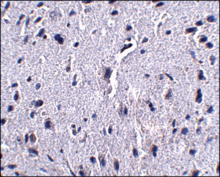
Immunohistochemistry Analysis: A 1:250 dilution of this antibody detected PD-1 in human tonsil and human bone marrow tissues.
Immunohistochemistry Analysis: A 1:250 dilution of this antibody detected PD-1 in human tonsil and human bone marrow tissues.
標靶描述
31.65 kDa calculated.
外觀
Protein G purified
Format: Purified
Purified mouse monoclonal antibody IgG2a in buffer containing 0.1 M Tris-Glycine (pH 7.4), 150 mM NaCl with 0.05% sodium azide.
儲存和穩定性
Stable for 1 year at 2-8°C from date of receipt.
其他說明
Concentration: Please refer to lot specific datasheet.
免責聲明
Unless otherwise stated in our catalog or other company documentation accompanying the product(s), our products are intended for research use only and are not to be used for any other purpose, which includes but is not limited to, unauthorized commercial uses, in vitro diagnostic uses, ex vivo or in vivo therapeutic uses or any type of consumption or application to humans or animals.
未找到合适的产品?
试试我们的产品选型工具.
儲存類別代碼
12 - Non Combustible Liquids
水污染物質分類(WGK)
WGK 1
我们的科学家团队拥有各种研究领域经验,包括生命科学、材料科学、化学合成、色谱、分析及许多其他领域.
联系技术服务部门


