推荐产品
生物源
rabbit
品質等級
共軛
ALEXA FLUOR™ 488
抗體表格
affinity isolated antibody
抗體產品種類
primary antibodies
無性繁殖
polyclonal
物種活性
rat, mouse
物種活性(以同源性預測)
human (based on 100% sequence homology)
技術
immunocytochemistry: suitable
immunohistochemistry: suitable
NCBI登錄號
UniProt登錄號
運輸包裝
wet ice
目標翻譯後修改
unmodified
基因資訊
human ... TUBB3(10381)
mouse ... Tubb3(22152)
rat ... Tubb3(246118)
一般說明
微管蛋白是小球状蛋白家族的几个成员之一,这些蛋白聚合形成称为微管的细胞骨架结构。微管蛋白家族中最常见的成员是α-微管蛋白和β-微管蛋白。通常由α-和β-微管蛋白亚基组成的微管蛋白二聚体组装到形成大聚合物的微管上。β-III微管蛋白是一种独特的微管亚基,几乎只在神经元中表达。由于β-III微管蛋白的独特表达模式,它通常被用作标记物,以从不表达它的其他脑细胞(例如神经胶质)中识别神经元细胞。β-III微管蛋白也可作为细胞结构标记,因为它主要存在于神经突延伸内。
特異性
该抗体可识别βIII微管蛋白。
免疫原
对应于人βIII微管蛋白的合成线性肽。
應用
免疫组化:代表性皮层已显示在成年小鼠大脑中起作用。
研究子类别
神经丝&神经元代谢
神经丝&神经元代谢
研究类别
神经科学
神经科学
品質
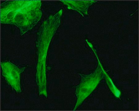
已通过免疫细胞化学在大鼠E18皮层细胞中进行评估。
免疫细胞化学分析:该抗体的1:400稀释液在大鼠E18皮质细胞中检测到βIII微管蛋白。
免疫细胞化学分析:该抗体的1:400稀释液在大鼠E18皮质细胞中检测到βIII微管蛋白。
標靶描述
计算分子量为50 kDa
外觀
亲和纯化
纯化的兔多克隆抗体与含0.1%叠氮化钠和15 mg/ml BSA的PBS中的Alexa Fluor™ 488偶联。
儲存和穩定性
自发运之日起,在 2-8°C 条件下可稳定保存1年
分析報告
对照
大鼠E18皮层细胞
大鼠E18皮层细胞
法律資訊
ALEXA FLUOR is a trademark of Life Technologies
免責聲明
除非我们的产品目录或产品附带的其他公司文档另有说明,否则我们的产品仅供研究使用,不得用于任何其他目的,包括但不限于未经授权的商业用途、体外诊断用途、离体或体内治疗用途或任何类型的消费或应用于人类或动物。
未找到合适的产品?
试试我们的产品选型工具.
儲存類別代碼
12 - Non Combustible Liquids
水污染物質分類(WGK)
WGK 2
閃點(°F)
Not applicable
閃點(°C)
Not applicable
其他客户在看
Predrag Kalaba et al.
Biomolecules, 13(9) (2023-09-28)
The high structural similarity, especially in transmembrane regions, of dopamine, norepinephrine, and serotonin transporters, as well as the lack of all crystal structures of human isoforms, make the specific targeting of individual transporters rather challenging. Ligand design itself is also
Jianzhang Hu et al.
Investigative ophthalmology & visual science, 60(1), 16-25 (2019-01-03)
To investigate the contribution and mechanism of miRNAs and autophagy in diabetic peripheral neuropathy. In this study, we used streptozotocin (STZ)-induced type I diabetes C57 mice as animal models, and we detected the expression of miR-34c and autophagic intensity in
Zhenzhen Zhang et al.
Molecular vision, 24, 274-285 (2018-04-13)
To investigate the effect and mechanism of proresolving lipid mediator resolvin D1 (RvD1) on the corneal epithelium and the restoration of mechanical sensation in diabetic mice. Type 1 diabetes was induced in mice with intraperitoneal streptozocin injections. The healthy and
Emily R Aurand et al.
Stem cell research, 12(1), 11-23 (2013-10-22)
Hydrogels provide a unique tool for neural tissue engineering. These materials can be customized for certain functions, i.e. to provide cell/drug delivery or act as a physical scaffold. Unfortunately, hydrogel complexities can negatively impact their biocompatibility, resulting in unintended consequences.
Yuan Zhang et al.
Frontiers in molecular biosciences, 8, 737472-737472 (2021-09-14)
Diabetes mellitus (DM) is a complex metabolic disorder. Long-term hyperglycemia may induce diabetic keratopathy (DK), which is mainly characterized by delayed corneal epithelial regeneration. MicroRNAs (miRNAs) have been reported to play regulatory roles during tissue regeneration. However, the molecular mechanism
我们的科学家团队拥有各种研究领域经验,包括生命科学、材料科学、化学合成、色谱、分析及许多其他领域.
联系技术服务部门


