Skip to Content
Merck
All Photos(1)

Key Documents

358R-1

Sigma-Aldrich

MUM1 (EP190) Rabbit Monoclonal Primary Antibody

Synonym(s):

Multiple Myeloma Oncogene-1

Sign Into View Organizational & Contract Pricing


About This Item

UNSPSC Code:
12352203

biological source

rabbit

Quality Level

100
500

conjugate

unconjugated

antibody form

culture supernatant

antibody product type

primary antibodies

clone

EP190, monoclonal

description

For In Vitro Diagnostic Use in Select Regions

form

buffered aqueous solution

species reactivity

human

packaging

vial of 0.1 mL concentrate (358R-14)
vial of 0.1 mL concentrate Research Use Only (358R-14-RUO)
vial of 0.5 mL concentrate (358R-15)
vial of 1.0 mL concentrate (358R-16)
vial of 1.0 mL concentrate Research Use Only (358R-16-RUO)
vial of 1.0 mL pre-dilute Research Use Only (358R-17-RUO)
vial of 1.0 mL pre-dilute ready-to-use (358R-17)
vial of 7.0 mL pre-dilute ready-to-use (358R-18)
vial of 7.0 mL pre-dilute ready-to-use Research Use Only (358R-18-RUO)

manufacturer/tradename

Cell Marque

technique(s)

immunohistochemistry (formalin-fixed, paraffin-embedded sections): 1:100-1:500 (concentrated)

isotype

IgG

control

diffuse large B-cell (lymphoma), plasma cell myeloma (tumor), tonsil

shipped in

wet ice

storage temp.

2-8°C

visualization

cytoplasmic, nuclear

Gene Information

human ... PWWP3A(84939)

General description

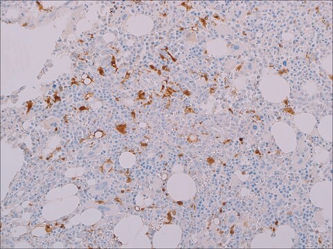
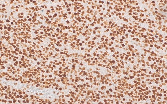
Anti-MUM1 antibody labels a 50kDa, multiple myeloma oncogene-1 (MUM1) protein. MUM1 is encoded by the MUM1/IRF-4 gene, which is mapped to 6q23-25 and identified as a myeloma-associated oncogene. It is a member of the interferon regulatory factor family of transcription factors and plays an important role in the regulation of gene expression in response to signaling by interferon and other cytokines. MUM1 positive cells express the protein in the nucleus in a diffuse and microgranular pattern. However, some positivity is also observed in the cytoplasm of MUM1-expressing cells. In normal/reactive lymphoid tissues, such as lymph node, this antibody stains plasma cells, some B-cells in the light zone of germinal centers, and a subset of T-cells (T-cells in germinal centers and interfollicular areas). MUM1 expression has been described in diffuse large B-cell lymphoma (DLBCL). Anti-MUM1 antibody can stain other B-cell lymphomas such as lymphoplasmacytic lymphoma, chronic lymphocytic leukemia, follicular lymphoma, marginal zone lymphoma, lymphoblastic lymphoma/leukemia, primary effusion lymphoma, DLBCL, Burkitt-like lymphoma, and classical Hodgkin lymphoma. However, the tumor cells in nodular lymphocyte predominant Hodgkin lymphoma are negative or only weakly positive. MUM1 is also expressed in plasma cell myeloma.

Quality


IVD

IVD

IVD

RUO

Linkage

MUM1 Positive Control Slides, Product No. 358S, are available for immunohistochemistry (formalin-fixed, paraffin-embedded sections).

Physical form

Solution in Tris Buffer, pH 7.3-7.7, with 1% BSA and <0.1% Sodium Azide.

Preparation Note

Download the IFU specific to your product lot and format
Note: This requires a keycode which can be found on your packaging or product label.

Download the latest released IFU
Note: This IFU may not apply to your specific product lot.

Other Notes

For Technical Service please contact: 800-665-7284 or email: service@cellmarque.com

Legal Information

Cell Marque is a trademark of Merck KGaA, Darmstadt, Germany

Not finding the right product?  

Try our Product Selector Tool.

Storage Class Code

12 - Non Combustible Liquids

WGK

WGK 2

Flash Point(F)

Not applicable

Flash Point(C)

Not applicable


Certificates of Analysis (COA)

Search for Certificates of Analysis (COA) by entering the products Lot/Batch Number. Lot and Batch Numbers can be found on a product’s label following the words ‘Lot’ or ‘Batch’.

Already Own This Product?

Find documentation for the products that you have recently purchased in the Document Library.

Visit the Document Library

T Yamagata et al.
Molecular and cellular biology, 16(4), 1283-1294 (1996-04-01)
We have isolated a novel cDNA clone encoding interferon (IFN) consensus sequence-binding protein in adult T-cell leukemia cell line or activated T cells (ICSAT); this protein is the human homolog of the recently cloned Pip/LSIRF. ICSAT is structurally most closely
W Kempf et al.
The British journal of dermatology, 158(6), 1280-1287 (2008-04-16)
Primary cutaneous CD30+ lymphoproliferative disorders include lymphomatoid papulosis (LyP) and primary cutaneous CD30+ anaplastic large T-cell lymphoma (ALCL). Because of overlapping histological features, it is impossible to distinguish ALCL from LyP on histological grounds. MUM1 (Multiple Myeloma oncogene 1) is
Gabriela Gualco et al.
Human pathology, 40(4), 565-571 (2009-01-16)
Burkitt lymphoma is a highly aggressive non-Hodgkin lymphoma with endemic, sporadic, and immunodeficiency-associated clinical variants composed of monomorphic medium-sized B cells with a high proliferation rate and a translocation involving the C-MYC locus. Classically, the immunophenotype of Burkitt lymphoma has
Uma Sundram et al.
Modern pathology : an official journal of the United States and Canadian Academy of Pathology, Inc, 16(8), 802-810 (2003-08-16)
The diagnosis of malignant melanoma remains one of the most difficult to render in surgical pathology, partially because of its extreme histologic variability. Limits in the sensitivity and/or specificity of the currently available melanocytic markers such as anti-S100, HMB45, and
Christine P Hans et al.
Blood, 103(1), 275-282 (2003-09-25)
Diffuse large B-cell lymphoma (DLBCL) can be divided into prognostically important subgroups with germinal center B-cell-like (GCB), activated B-cell-like (ABC), and type 3 gene expression profiles using a cDNA microarray. Tissue microarray (TMA) blocks were created from 152 cases of

Our team of scientists has experience in all areas of research including Life Science, Material Science, Chemical Synthesis, Chromatography, Analytical and many others.

Contact Technical Service