Skip to Content
Merck
All Photos(2)

Key Documents

05-719

Sigma-Aldrich

Anti-Phosphatidylserine Antibody, clone 1H6

clone 1H6, Upstate®, from mouse

Synonym(s):

Anti-Phosphatidylserine, Clone 1H6 Anti-Phosphatidylserine, Phosphatidylserine Detection

Sign Into View Organizational & Contract Pricing


About This Item

UNSPSC Code:
12352203
eCl@ss:
32160702
NACRES:
NA.41

biological source

mouse

Quality Level

antibody form

purified immunoglobulin

antibody product type

primary antibodies

clone

1H6, monoclonal

species reactivity

vertebrates

manufacturer/tradename

Upstate®

technique(s)

flow cytometry: suitable
immunohistochemistry: suitable

isotype

IgG

shipped in

dry ice

target post-translational modification

phosphorylation (pSer)

Gene Information

human ... PTDSS1(9791)

General description

Significance:
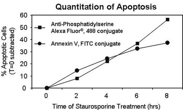
Anti-Phosphatidylserine may be used to detect translocation of the membrane phospholipid phosphatidylserine (PS) from the inner to the outer cell membrane leaflet; it provides an alternative to Annexin V for quantitation of apoptosis.
Phosphatidylserine (PS) is a phospholipid (1,2-diacyl-sn-glycerol-(3)-L-phosphoserine) found in the membrane of all plants and animals. PS is most concentrated in the brain, as PS makes up about 10% of the lipid content of neurons. Neural PS is thought to play a role in maintaining nerve cell integrity, enhancing neurotransmitter signal efficiency, and improving nerve cell signal transmission. One of the key features of apoptosis is the expresion of PS on the outer cell membrane. PS is usually found on the inner side of the plasma membrane, but during early stage apoptosis, the PS is translocated to the outer membrane. FITC-labeled annexin V can bind to the exposed PS, providing the basis of most annexin V-based apoptosis assays.

Specificity

Recognizes phosphatidylserine (PS) in cell membranes

Immunogen

Liposomes containing 70% phosphatidylserine and 30% phosphatidylglycerol

Application

Anti-Phosphatidylserine Antibody, clone 1H6 is an antibody against Phosphatidylserine for use in FC, IH.
Flow Cytometry: 0.2-2 μg/mL of a previous lot detected cell surface phosphatidylserine on Jurkat cells treated with Fas Ligand vesicles (Catalog # 01-210). See page two.

Immunohistochemistry: This antibody has been reported by an independent laboratory to detect phosphatidylserine in rat carotid artery paraffin sections fixed with ethanol/acetone (Mandinov, L., 2003).
Research Category
Apoptosis & Cancer
Research Sub Category
Apoptosis - Additional

Quality

routinely evaluated by flow cytometry on Jurkat cells treated with Fas Ligand vesicles (Catalog #01-210)

Target description

none given

Physical form

Format: Purified
Protein G Purified
Purified IgG supplied in 0.1M Tris-Glycine, 0.15M NaCl, 0.05% Sodium Azide, pH 7.4 before the addition of glycerol to 30%. Liquid at -20°C.

Storage and Stability

Stable for 1 year at from date of receipt.

Analysis Note

Control
Brain tissue

Other Notes

Concentration: Please refer to the Certificate of Analysis for the lot-specific concentration.

Legal Information

UPSTATE is a registered trademark of Merck KGaA, Darmstadt, Germany

Disclaimer

Unless otherwise stated in our catalog or other company documentation accompanying the product(s), our products are intended for research use only and are not to be used for any other purpose, which includes but is not limited to, unauthorized commercial uses, in vitro diagnostic uses, ex vivo or in vivo therapeutic uses or any type of consumption or application to humans or animals.

Not finding the right product?  

Try our Product Selector Tool.

Storage Class Code

10 - Combustible liquids

WGK

WGK 1


Certificates of Analysis (COA)

Search for Certificates of Analysis (COA) by entering the products Lot/Batch Number. Lot and Batch Numbers can be found on a product’s label following the words ‘Lot’ or ‘Batch’.

Already Own This Product?

Find documentation for the products that you have recently purchased in the Document Library.

Visit the Document Library

Jiunn-Min Shieh et al.
International journal of medical sciences, 10(8), 988-994 (2013-06-27)
Previously, we identified a sequence variant (N375S) of c-Met gene, however, its association with lung cancer risk and prognosis remain undefined. We investigated the genotype distribution of the c-Met-N375S sequence variant in 206 lung cancer patients and 207 non-cancer controls
Phosphatidylserine receptor and apoptosis: consequences of a non-ingested meal.
Botto, Marina
Arthritis Research & Therapy, 6, 147-150 (2004)
I Vermes et al.
Journal of immunological methods, 184(1), 39-51 (1995-07-17)
In the early stages of apoptosis changes occur at the cell surface, which until now have remained difficult to recognize. One of these plasma membrane alterations is the translocation of phosphatidylserine (PS) from the inner side of the plasma membrane
Manjarika De et al.
Molecular therapy. Nucleic acids, 10, 9-27 (2018-03-04)
There is a pressing need for a ubiquitously expressed antigen or receptor on the tumor surface for successful mitigation of the deleterious side effects of chemotherapy. Phosphatidylserine (PS), normally constrained to the intracellular surface, is exposed on the external surface
The role of phosphatidylserine in recognition and removal of erythrocytes.
Kuypers, F A and de Jong, K
Cellular and molecular biology (Noisy-le-Grand, France), 50, 147-158 (2004)

Our team of scientists has experience in all areas of research including Life Science, Material Science, Chemical Synthesis, Chromatography, Analytical and many others.

Contact Technical Service