Skip to Content
Merck
All Photos(1)

Documents

A2543

Sigma-Aldrich

Anti-α-Actinin antibody produced in rabbit

whole antiserum

Synonym(s):

Anti-CMD1AA, Anti-CMH23, Anti-MPD6, Anti-MYOCOZ

Sign Into View Organizational & Contract Pricing


About This Item

MDL number:
UNSPSC Code:
12352203
NACRES:
NA.41

biological source

rabbit

conjugate

unconjugated

antibody form

whole antiserum

antibody product type

primary antibodies

clone

polyclonal

contains

15 mM sodium azide

species reactivity

chicken

technique(s)

indirect immunofluorescence: 1:500 using cultured chicken fibroblasts
western blot: suitable using cultured cells and chicken gizzard extract

UniProt accession no.

shipped in

dry ice

storage temp.

−20°C

target post-translational modification

unmodified

Gene Information

General description

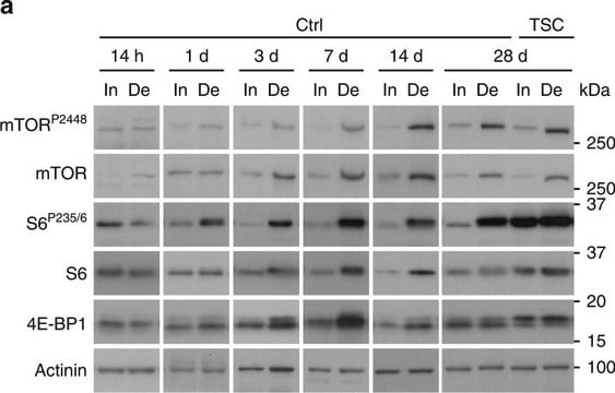
α-Actinin is a 100kDa actin-binding protein that is found in muscle as well as non-muscle cells. Rabbit anti-α-actinin antibody stains α-actinin in cell cultures using indirect immunofluorescence. The antibody reacts best with cultured chicken fibroblasts, however, adequate labeling may also be obtained with cells of other species. In an immunoblot assay, the antiserum specifically stains the α-actinin band.
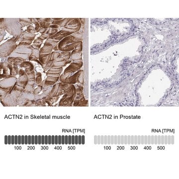
α-Actinin is a member of the spectrin superfamily. It consists of an antiparallel homodimer, with an N-terminal actin-binding domain (ABD), a central domain of four spectrin-like repeats (SRs), and a C-terminal calmodulin-like domain (CAMD) with two pairs of EF-hand motifs (EFs). α-Actinin exists in four isoforms: α-actinin 1, α-actinin 2, α-actinin 3 and α-actinin 4. In smooth muscles, α-actinin is present in dense bodies and plaques whereas, in normal skeletal muscles, it is associated with Z-discs that define muscle sarcomere.

Specificity

Anti-α-Actinin specifically stains α-actinin in cell cultures using indirect immunfluorescent labeling techniques. The antibody reacts best with cultured chicken fibroblasts, however adequate labeling may also be obtained with cells of other species.

Immunogen

α-actinin from chicken gizzard

Application

Anti-Actinin antibody was used to detect actinin in human mesangial cells. Cells were first grown on glass cover slips, fixed in cold methanol and permeabilized with 3.5% paraformaldehyde/0.1% Tween-20 for 5 min at room temperature.
Rabbit anti-α-actinin antibody has been used for:
  • co-immunoprecipitation studies
  • immunoblotting studies
  • immunofluorescence

Biochem/physiol Actions

α-Actinin is an actin-crosslinking Z-disk protein. It plays a vital role in linking the cytoskeleton to different transmembrane proteins in a variety of junctions. α-Actinin modulates receptor activities and serves as a cytoskeleton scaffold for diverse signaling pathways. It is implicated in muscle contraction.

Physical form

Supplied as a liquid with 0.1% sodium azide as a preservative.

Storage and Stability

For continuous use, store at 2-8 °C for up to one month. For extended storage, the solution may be frozen in working aliquots. Repeated freezing and thawing is not recommended. Storage in "frost-free" freezers not recommended. If slight turbidity occurs upon prolonged storage, clarify the solution by centrifugation before use.

Disclaimer

Unless otherwise stated in our catalog or other company documentation accompanying the product(s), our products are intended for research use only and are not to be used for any other purpose, which includes but is not limited to, unauthorized commercial uses, in vitro diagnostic uses, ex vivo or in vivo therapeutic uses or any type of consumption or application to humans or animals.

Not finding the right product?  

Try our Product Selector Tool.

Storage Class Code

10 - Combustible liquids

WGK

WGK 3

Flash Point(F)

Not applicable

Flash Point(C)

Not applicable


Certificates of Analysis (COA)

Search for Certificates of Analysis (COA) by entering the products Lot/Batch Number. Lot and Batch Numbers can be found on a product’s label following the words ‘Lot’ or ‘Batch’.

Already Own This Product?

Find documentation for the products that you have recently purchased in the Document Library.

Visit the Document Library

B Sjöblom et al.
Cellular and molecular life sciences : CMLS, 65(17), 2688-2701 (2008-05-20)
Alpha-actinin is a cytoskeletal actin-binding protein and a member of the spectrin superfamily, which comprises spectrin, dystrophin and their homologues and isoforms. It forms an anti-parallel rod-shaped dimer with one actin-binding domain at each end of the rod and bundles
Feifei Su et al.
Molecular medicine reports, 23(3) (2021-01-27)
High‑mobility group box 1 (HMGB1) is released by necrotic cells and serves an important role in cardiovascular pathology. However, the effects of HMGB1 in cardiomyocyte hypertrophy remain unclear. Therefore, the aim of the present study was to investigate the potential role
Ana Virel et al.
Molecular biology and evolution, 21(6), 1024-1031 (2004-03-12)
The N-terminal actin-binding domain of alpha-actinin is connected to the C-terminal EF-hands by a rod domain. Because of its ability to form dimers, alpha-actinin can cross-link actin filaments in muscle cells as well as in nonmuscle cells. In the prototypic
Manuel Ramos-Kuri et al.
Biochimica et biophysica acta, 1853(11 Pt A), 2870-2884 (2015-08-12)
The importance of the oncogene Ras in cardiac hypertrophy is well appreciated. The hypertrophic effects of the constitutively active mutant Ras-Val12 are revealed by clinical syndromes due to the Ras mutations and experimental studies. We examined the possible anti-hypertrophic effect
L J Swatzell et al.
Plant & cell physiology, 40(2), 173-183 (1999-04-15)
Integrins are a large family of integral membrane proteins that function in signal transduction in animal systems. These proteins are conserved in vertebrates, invertebrates, and fungi. Evidence from previous research suggests that integrin-like proteins may be present in plants as

Our team of scientists has experience in all areas of research including Life Science, Material Science, Chemical Synthesis, Chromatography, Analytical and many others.

Contact Technical Service