General description
ColorWheel® technology is a novel and proprietary method of creating your own antibody and dye combinations for use in flow cytometry. By incubating any ColorWheel® antibody with any ColorWheel® dye, researchers can quickly and simply produce primary conjugated antibodies for use in single color or multicolor flow cytometry analysis. Each ColorWheel® antibody and dye is lyophilized for long-term storage, allowing for simplicity and flexibility without compromise. ColorWheel® technology requires both ColorWheel® antibody and ColorWheel® dye for Flow Cytometry application.
We are committed to bringing you greener alternative products, which adhere to one or more of The 12 Principles of Green Chemistry.This product is Preservative-free, lyophilized product for enhanced stability and allow for ambient shipping and thus aligns with "Waste Prevention", "Designing Safer Chemicals" and "Design for Energy Efficiency".
Click here for more information.
Specificity
Clone 9F10 is a mouse monoclonal antibody that detects CD49d (Integrin a-4).
Application
Quality Control Testing
Evaluated by Flow Cytometry in Human peripheral blood mononuclear cells (PBMC).
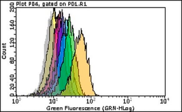
Flow Cytometry Analysis: Staining of one million Human peripheral blood mononuclear cells (PBMC) was performed using 5 μL of a 1:1 mixture of Cat. No. CWA-1086, Anti-Human CD49d / ITGA4 (9F10) ColorWheel® Dye-Ready mAb and Cat. No. CWDS-PE ColorWheel® Antibody-Ready Phycoerythrin (PE) Dye or an equivalent amount of PE-conjugated Mouse IgG1 isotype control.
Note: Actual optimal working dilutions must be determined by end user as specimens, and experimental conditions may vary with the end user
Compatibility
Requires ColorWheel® Dye (Sold Separately)
Target description
Integrin alpha-4 (UniProt: P13612; also known as CD49 antigen-like family member D, Integrin alpha-IV, VLA-4 subunit alpha, CD49d) is encoded by the ITGA4 (also known as CD49D) gene (Gene ID: 3676) in human. Integrins are heterodimers composed of a variable a-subunit of 150-170 kDa and a conserved 95 kDa b subunit. They contain a large extracellular domain responsible for ligand binding, a single transmembrane domain, and a cytoplasmic domain. The exact combination of various a- and b-subunits dictates the binding specificity of integrins to different ECM components. Although both subunits are required for adhesion, the binding specificity primarily depends on the extracellular portion of the a-subunit. Integrins bind to their ligands with a low affinity and this binding occurs only when a certain minimal number of integrins are present at specific points known as focal contacts and hemidesmosomes. In response to specific stimuli, they cluster in focal contacts and their combined affinities create a region on the cell surface, which presents sufficient adhesive capacity to adhere to the ECM. This allows cells to bind to a large numbers of matrix molecules simultaneously while still maintaining their ability to explore their environment without losing all attachmentsCD49d is a single-pass type I membrane glycoprotein that is synthesized with a signal peptide (aa 1-33), which is subsequently cleaved off to produce the mature form that contains a large extracellular domain (aa 34-977), a transmembrane domain (aa 978-1001), and a short cytoplasmic tail (aa 1002-1032). CD49d plays a critical role in leucocyte trafficking, activation, and survival, and also facilitates interactions between leucocytes and stromal cells found in the marrow or germinal center of lymphoid follicles via VCAM-1 and fibronectin. It also serves as a signalling receptor that influences B-cell survival via upregulation of Bcl-2 family members. CD49d/CD29 and CD49d/Integrin 7 are known to be receptors for fibronectin. They recognize one or more domains within the alternatively spliced CS-1 and CS-5 regions of fibronectin. They also serve as receptors for VCAM1. CD40d/CD29 recognizes the sequence Q-I-D-S in VCAM1. (Ref.: Haile, LA., et al. (2010). J. Immunol. 185; 203-210; Shanafelt, TD., et al. (2008). Br. J. Haematol. 140(5); 537-546).
Physical form
Lyophilized from PBS containing D-Mannitol and Sucrose, normal appearance is a dried pellet. Reconstituted antibody solution is stable and functional as assessed by functional testing. Contains no biocide or preservatives, such as azide.
Reconstitution
1.0 mg/mL after reconstitution at 25 μL in PBS. Please refer to guidance on suggested starting dilutions and/or titers per application and sample type.
Storage and Stability
Recommend storage of lyophilized product at 2-8°C. Before reconstitution, micro-centrifuge vials briefly to spin down material to bottom of the vial. Reconstitute each vial by adding 25 μL PBS. Protect from light.
Legal Information
ColorWheel is a registered trademark of Merck KGaA, Darmstadt, Germany
Disclaimer
Unless otherwise stated in our catalog or other company documentation accompanying the product(s), our products are intended for research use only and are not to be used for any other purpose, which includes but is not limited to, unauthorized commercial uses, in vitro diagnostic uses, ex vivo or in vivo therapeutic uses or any type of consumption or application to humans or animals.


