Metabolites – A Serious Challenge for LC/MS Separations and Usual Detection Techniques
Rudi Köhling
Analytix 5, 2010
Although the genome of several organisms has been decoded, the knowledge about the expressed proteins, and their complex interactions and catalyzed chemical reactions remains incomplete. Metabolomics encompasses the study of all compounds and reaction products taking part in and regulating these cellular processes. Metabolomics may be applied to a single cell or an organism as a whole.1
Biosynthesis of Cholesterol and Fatty-Acids (Biofuels)
Cholesterol is an important molecule in the human body. It is incorporated in cell membranes and stabilizes the lipid bi-layers. Modifications to this molecule by other enzymes yield another important group of compounds: steroid hormones. These hormones assume a role in signal transduction and control the protein biosynthesis; for example, estrogens such as estradiol are responsible for the growth of female genitals and regulate the menstruation. Surprisingly, the cholesterol building blocks also take part in the biosynthesis of fatty-acids, which can replace mineral oil-based fuels.2
Figure 1 displays some of the steps of the pathway leading to the formation of cholesterol. In a large number of these reactions, a phosphorylation of hydroxyl moieties with ATP takes place; which in turn means that very hydrophilic compounds coexist with hydrophobic compounds, and a separation by chromatography places varying demands on the different stationary phases.
The example above provides only a small insight into the innumerable reactions with thousands of reaction products and intermediates. Some of these metabolites have pathological relevance and refer to several different disease patterns, e. g. phenylketonuria (PKU).
We offer the tools and the knowledge for the study of metabolic processes and the analysis of the metabolites. A large number of certified reference standards, enzymes, substrates, and chromatographic products can be found on our website.

Figure 1.Biosynthesis of IPP and DMAP via mevalonate pathway. These isoprenoid phosphates are the smallest units involved in the production of a large number of vitamins, coenzymes, hormones, lipids.
Analysis of Metabolites
NMR, LC/MS, CE(/MS) and GC/MS are typically the methods of choice to separate, identify and quantify in the analysis of biological samples. However, most of the metabolites, especially those listed in Table 1, are very polar and do not show any UV absorption, which excludes the use of UV or fluorescence detectors for HPLC. Sample size: approx. 0.3 g. The sample can be easily handled using a syringe without a needle.
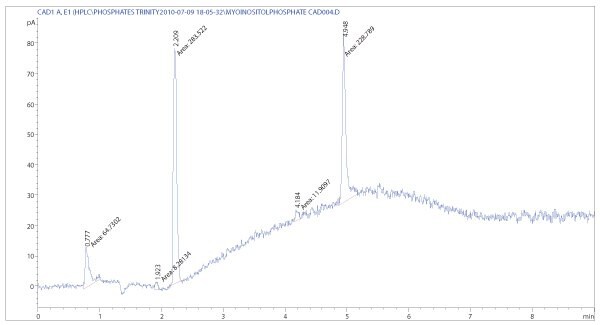
The Corona CAD represents a new detection technique which closes the gap between optical and mass detectors and offers both sensitivity and responses, depending on the concentration of the analyte. It utilizes ionization processes comparable to APCI sources and detects charged particles. The particles are formed by drying the mobile phase with nitrogen, which is also used to charge the particles by passing a corona discharge needle. These detectors have gained increasing importance in analytical laboratories because of their ease of use, low price and high sensitivity compared to other detectors, e. g. refractive index. They can be used for controlling the quality, purity and content of pure metabolites (Figures 2 and 3).

Figure 2.CAD chromatogram of myo-ionisitol triphosphate sodium salt. One special feature of this detector is the ability to detect alkali cations as well (2.2 min). Mobile phase compositions can be transferred from LC/MS methods as the Corona CAD also depends on volatile additives.

Figure 3.Separation of myo-inositol phosphates by HILIC. The method is suitable for MS and CAD detection. Best peak shapes can be obtained at a higher pH.
Liquid Chromatography
The separation of such polar compounds by HPLC poses a challenge for the method development. Ion exchange, HILIC, or some chiral stationary phases (e. g. Chirobiotic™/ Cyclobond columns), may be a good starting point for LC method development using MS or CAD detection. High buffer concentrations and varying pH conditions influence retention and peak shape crucially, which is useful for the optimization. Myo-inositol tri- and -pentaphosphates can be separated by HILIC using a Supelco® Ascentis® Express HILIC column. Figure 3 shows the extracted ion chromatograms of the [M+H]+ ions.
Another more difficult separation is related to the isoprenoid phosphate compounds in Figure 1. Isopentenyl phosphate (IP), dimethylallyl phosphate (DMAP), and their related pyrophosphates, IPP and DMAPP, differ only in the position of the double bond. However, the HPLC separation is possible with a Supelco Cyclobond I 2000 stationary phase (Figure 4).
Finally, the separation of OPA-derivatized amino acids is presented in Figure 5. There are additional methods for the analysis of amino acids by HPLC described in the literature.3-5 Most of these techniques use pre-column derivatization to change the polarity of the analytes, and to make them detectable for UV and fluorescence detectors. Unfortunately, additional peaks of the derivatization agent may overlay with analyte peaks; however, this problem can be avoided by using guard columns and switching between various wave lengths.
The OPA/FMOC reaction4 can easily be transferred to Ascentis Express C8/C18 columns; in particular, standard HPLC systems profit from their very high resolution. Only a 5 cm column is necessary to separate all 16 amino acid derivatives within a run-time of 10 min (Agilent® 1200 system).
The various methods and techniques used for metabolomics all require the use of reference standards. We have introduced a new quality of organic TraceCERT® standards, beginning with a series of amino acids, which are used as external standards for the determination of amino acid concentrations in various matrices. Amino acids are also important biomarkers for some hereditary metabolic diseases and can be detected within a prenatal/neo- natal screening.1,5 The precision of the analysis strongly depends upon the exact knowledge of the analyte content in the reference standards. This is guaranteed by using high-precision qNMR measurements, and the production of such standards in an accredited laboratory (see previous article by J. Wüthrich).

Figure 4.Separation of IP, DMAP, IPP and DMAPP by chiral chromatography with a cyclodextrin phase (Cyclobond I 2000).

Figure 5.Separation of 15 amino acids (OPA derivatives) using pre-column derivatization and reversed-phase chromatography (Ascentis Express C8 column) according to. All amino acid derivatives, as well as the agents, are separated very well. The peak of the OPA reagent can be suppressed by using another wave length.
References
계속 읽으시려면 로그인하거나 계정을 생성하세요.
계정이 없으십니까?