おすすめの製品
由来生物
mouse
品質水準
結合体
unconjugated
抗体製品の状態
purified antibody
抗体製品タイプ
primary antibodies
クローン
7B10-3E4, monoclonal
分子量
calculated mol wt 45.63 kDa
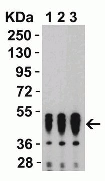
observed mol wt ~50 kDa
化学種の反応性
virus
包装
antibody small pack of 100 μg
テクニック
ELISA: suitable
immunofluorescence: suitable
immunoprecipitation (IP): suitable
western blot: suitable
アイソタイプ
IgG1κ
UniProtアクセッション番号
輸送温度
dry ice
保管温度
2-8°C
ターゲットの翻訳後修飾
unmodified
詳細
SARS-CoV-2 Nucleoprotein (UniProt: P0DTC9; also known as N, Nucleocapsid protein, NC, Protein N) is encoded by the N gene (Gene ID: 43740575) in SARS-CoV-2 virus. The SARS-CoV-2 is a positive-strand RNA virus that causes severe respiratory syndrome in human. The mature SARS-CoV-2 contains 4 structural proteins: Envelope (E), Membrane (M), Nucleocapsid (N), and the Spike protein (S). E and M proteins help in viral assembly and N protein is needed for RNA synthesis. The S protein is a single-pass type I, homotrimeric, membrane glycoprotein that is responsible for virus binding and entry into host cell. The N protein packages the positive strand viral genome RNA into a helical ribonucleocapsid (RNP) and plays a fundamental role during virion assembly through its interactions with the viral genome and membrane protein M. It plays an important role in enhancing the efficiency of subgenomic viral RNA transcription as well as viral replication. The RNA binding region of SARS-CoV-2 is localized to amino acids 41-186 and its dimerization domain is in amino acids 258-361. Lack of cysteine is one of the common features in nucleoprotein from different coronaviruses. The complete N protein of SARS-CoV and COV-2 is a highly basic with positive net charges and high pI (10.11). Its N-terminal region is more basic with positive charge, while the C-terminal region is acidic with negative charge. The middle region of nucleoprotein displays relatively higher hydrophobicity and the two termini display greater hydrophilicity. Nucleoprotein can undergo phosphorylation at multiple serines and threonines and some of these phosphorylation events are essential for RNA binding, oligomerization and localization to nucleoli. Phosphorylation is also required for recruitment of host RNA helicase DDX1 (DEAD-Box Helicase 1) that facilitates template readthrough and enables longer subgenomic mRNA synthesis. Clone 7B10-3E4 exhibits high reactivity with SARS-CoV-2 and weak reactivity with SARS-CoV-1.
特異性
Clone 7B10-3E4 is a mouse monoclonal antibody that detects nucleocapsid protein of Severe acute respiratory syndrome coronavirus 2 (SARS-CoV-2). It targets an epitope within the N-terminal domain. It also displays weak reactivity with SARS-CoV-1.
免疫原
His-tagged full-length nucleocapsid protein from Severe acute respiratory syndrome coronavirus 2 (SARS-CoV-2).
アプリケーション
Quality Control Testing
Evaluated by Western Blotting in lysates from HEK293T cells expressing SARS-CoV-2 Nucleocapsid protein.
Western Blotting Analysis (WB): A 1:1,000 dilution of this antibody detected Nucleocapsid protein in lysates from HEK293T cells expressing SARS-CoV-2 Nucleocapsid protein, but not in lysates from the wild type HEK293T cells.
Tested Applications
Western Blotting Analysis: A 1:20 dilution from a representative lot detected SARS-CoV-2 nucleocapsid protein in Whole cell lysates of HEK293T cells expressing the nucleocapsid (NC) proteins of various human coronaviruses (Courtesy of Stefan Schüchner, Ingrid Mudrak, Ingrid Frohner, and Egon Ogris (Max Perutz Labs, Medical University of Vienna, Austria).
ELISA Analysis: A 1:3 dilution from a representative lot detected SARS-CoV-2 nucleocapsid protein. (Courtesy of Stefan Schüchner, Ingrid Mudrak, Ingrid Frohner, and Egon Ogris (Max Perutz Labs, Medical University of Vienna, Austria).
Immunoprecipitation Analysis: A representative lot immunoprecipitated SARS-CoV-2 nucleocapsid protein. (Courtesy of Stefan Schüchner, Ingrid Mudrak, Ingrid Frohner, and Egon Ogris (Max Perutz Labs, Medical University of Vienna, Austria).
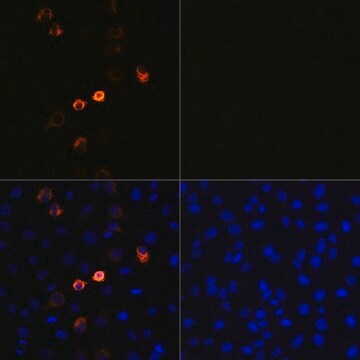
Immunofluorescence Analysis: A 1:5 dilution from a representative lot detected SARS-CoV-2 nucleocapsid in HeLa cells transiently transfected with SARS-CoV-2 NC-P2A-GFP (Courtesy of Stefan Schüchner, Ingrid Mudrak, Ingrid Frohner, and Egon Ogris (Max Perutz Labs, Medical University of Vienna, Austria).
Note: Actual optimal working dilutions must be determined by end user as specimens, and experimental conditions may vary with the end user
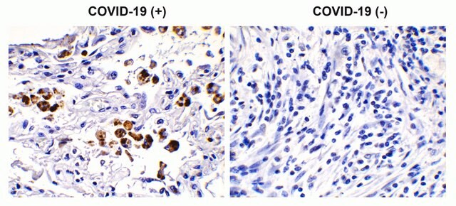
Anti-SARS-CoV-2 nucleocapsid, clone 7B10-3E4, Cat. No. MABF2788, is a mouse monoclonal antibody that detects SARS-CoV-2 Nucleoprotein and is tested for use in ELISA, Immunofluorescence, Immunoprecipitation, and Western Blotting.
物理的形状
Purified mouse monoclonal antibody IgG1 in buffer containing 0.1 M Tris-Glycine (pH 7.4), 150 mM NaCl, with 0.05% Sodium Azide without glycerol.
保管および安定性
Recommend storage at +2°C to +8°C. For long term storage antibodies can be kept at -20°C. Avoid repeated freeze-thaws.
その他情報
Concentration: Please refer to the Certificate of Analysis for the lot-specific concentration.
免責事項
Unless otherwise stated in our catalog or other company documentation accompanying the product(s), our products are intended for research use only and are not to be used for any other purpose, which includes but is not limited to, unauthorized commercial uses, in vitro diagnostic uses, ex vivo or in vivo therapeutic uses or any type of consumption or application to humans or animals.
Not finding the right product?
Try our 製品選択ツール.
保管分類コード
12 - Non Combustible Liquids
WGK
WGK 1
引火点(°F)
Not applicable
引火点(℃)
Not applicable
適用法令
試験研究用途を考慮した関連法令を主に挙げております。化学物質以外については、一部の情報のみ提供しています。 製品を安全かつ合法的に使用することは、使用者の義務です。最新情報により修正される場合があります。WEBの反映には時間を要することがあるため、適宜SDSをご参照ください。
Jan Code
MABF2788-100UG:
MABF2788-25UG:
試験成績書(COA)
製品のロット番号・バッチ番号を入力して、試験成績書(COA) を検索できます。ロット番号・バッチ番号は、製品ラベルに「Lot」または「Batch」に続いて記載されています。
ライフサイエンス、有機合成、材料科学、クロマトグラフィー、分析など、あらゆる分野の研究に経験のあるメンバーがおります。.
製品に関するお問い合わせはこちら(テクニカルサービス)
