
おすすめの製品


由来生物
mouse
品質水準
抗体製品の状態
purified immunoglobulin
抗体製品タイプ
primary antibodies
クローン
5C9/C8, monoclonal
化学種の反応性
human, mouse
テクニック
ChIP: suitable
immunocytochemistry: suitable
immunohistochemistry: suitable (paraffin)
western blot: suitable
アイソタイプ
IgG1κ
NCBIアクセッション番号
UniProtアクセッション番号
輸送温度
ambient
ターゲットの翻訳後修飾
unmodified
遺伝子情報
human ... TADA3(10474)
詳細
Transcriptional adapter 3 (UniProt O75528; also known as ADA3 homolog, ADA3-like protein, Alteration/deficiency in activation 3, hADA3, STAF54, Transcriptional adapter 3-like) is encoded by the TADA3 (also known as ADA3, TADA3L) gene (Gene ID 10474) in human. Alteration/deficiency in activation 3 (ADA3) is a component of several transcriptional co-activator and histone acetyltransferase (HAT) complexes and plays a critical role in in cell cycle regulation. Ada3 deletion in mice is embryonic lethal, and Ada3-deficient mouse embryonic fibroblasts (MEFs) exhiit a severe proliferation defect, dramatic changes in global histone acetylation, mitotic defects, as well as a delay in G2/M-to-G1 and G1-to-S transition. ADA3 also plays a role in genomic stability by controlling DNA repair checkpoints. ChIP-seq analsis reveals that ADA3 is significantly associated with human centromere regions across most chromosomes. In addition, ADA3 is found associated with CENP-B throughout all phases of the cell cycle, and CENP-B centromere binding decreased upon ADA3 knockdown. Wild-type human ADA3, but not CENP-B-binding deficient ADA3 mutant, prevented cell proliferation defects in MEFs following endogenous mouse ADA3 knockown. ADA3 overexpression and mislocalization correlates with poor prognosis in breast cancer patients.
特異性
Clone 5C9/C8 specifically detected Cre recombinase expression-induced ADA3 downregulation in Ada3FL/FL MEFs. Clone 5C9/C8 immunostained the nuclei of untransfected, but not ADA3 shRNA-transfected, 76N-TERT human mammary epithelial cells (Mohibi, S., et al. (2015). J. Biol. Chem. 290(47):28299-28310; Mohibi, S., et al. (2012). J. Biol. Chem. 287(35):29442-29456).
免疫原
Full-length recombinant human ADA3.
アプリケーション
Research Category
エピジェネティクス及び核内機能分子
エピジェネティクス及び核内機能分子
Anti-ADA3, clone 5C9/C8, Cat. No. MABE1057, is a highly specific mouse monoclonal antibody that targets TADA3 and has been tested in Chromatin Immunoprecipitation (ChIP), Immunocytochemistry, Immunohistochemistry (Paraffin), and Western Blotting.
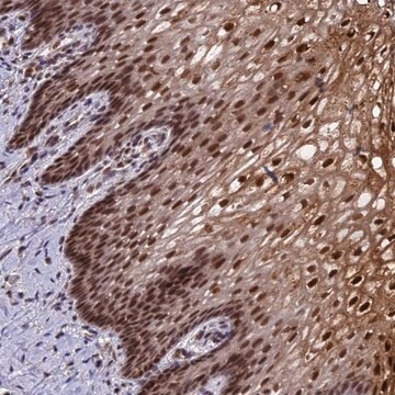
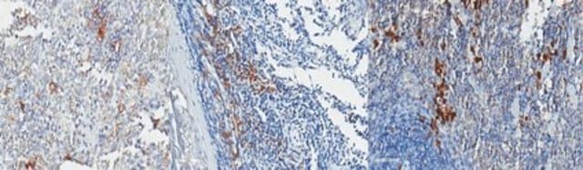
Immunohistochemistry Analysis: A representative lot detected nuclear ADA3 immunoreactivity in formalin-fixed, paraffin-embedded human breast carcinoma tissue section (Courtesy of Vimla Band, Ph.D., University of Nebraska USA).
Chromatin Immunoprecipitation (ChIP) Analysis: A representative lot detected ADA3 occupancy at the X chromosome centromere HOR region, the kinetochore assembly site also occupied by CENP-A and CENP-B, using TERT-immortalized human mammary epithelial cell 76N-TERT chromatin preparation. shRNA-mediated ADA3 knockdown led to a reduction in CENP-B, but not CENP-A, recruitment to the HOR region (Mohibi, S., et al. (2015). J. Biol. Chem. 290(47):28299-28310).
Immunocytochemistry Analysis: A representative lot detected ADA3 nuclear immunoreactivity in untransfected, but not ADA3 shRNA-transfected, TERT-immortalized human mammary epithelial 76N-TERT cells by fluorescent immunocytochemistry staining of 4% formaldehyde-fixed cells. Dual fluorescent staining revealed ADA3 and CENP-B nuclear colocalization (Mohibi, S., et al. (2015). J. Biol. Chem. 290(47):28299-28310).
Western Blotting Analysis: Representative lots detected both the endogenous mouse ADA3 (mADA3) and the exogenously expressed human ADA3 (hADA3) in Ada3FL/FL MEFs virally infected to express FLAG-tagged full-length or a.a. 111-432 hADA3 constructs. Cre recombinase expression downregulated only mADA3, but not hADA3 (Mohibi, S., et al. (2015). J. Biol. Chem. 290(47):28299-28310; Mohibi, S., et al. (2012). J. Biol. Chem. 287(35):29442-29456).
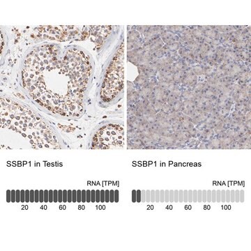
Western Blotting Analysis: A representative lot detected ADA3 expression levels among a panel of mouse tissues (Mohibi, S., et al. (2012). J. Biol. Chem. 287(35):29442-29456).
Chromatin Immunoprecipitation (ChIP) Analysis: A representative lot detected ADA3 occupancy at the X chromosome centromere HOR region, the kinetochore assembly site also occupied by CENP-A and CENP-B, using TERT-immortalized human mammary epithelial cell 76N-TERT chromatin preparation. shRNA-mediated ADA3 knockdown led to a reduction in CENP-B, but not CENP-A, recruitment to the HOR region (Mohibi, S., et al. (2015). J. Biol. Chem. 290(47):28299-28310).
Immunocytochemistry Analysis: A representative lot detected ADA3 nuclear immunoreactivity in untransfected, but not ADA3 shRNA-transfected, TERT-immortalized human mammary epithelial 76N-TERT cells by fluorescent immunocytochemistry staining of 4% formaldehyde-fixed cells. Dual fluorescent staining revealed ADA3 and CENP-B nuclear colocalization (Mohibi, S., et al. (2015). J. Biol. Chem. 290(47):28299-28310).
Western Blotting Analysis: Representative lots detected both the endogenous mouse ADA3 (mADA3) and the exogenously expressed human ADA3 (hADA3) in Ada3FL/FL MEFs virally infected to express FLAG-tagged full-length or a.a. 111-432 hADA3 constructs. Cre recombinase expression downregulated only mADA3, but not hADA3 (Mohibi, S., et al. (2015). J. Biol. Chem. 290(47):28299-28310; Mohibi, S., et al. (2012). J. Biol. Chem. 287(35):29442-29456).
Western Blotting Analysis: A representative lot detected ADA3 expression levels among a panel of mouse tissues (Mohibi, S., et al. (2012). J. Biol. Chem. 287(35):29442-29456).
品質
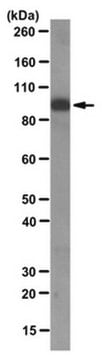
Evaluated by Western Blotting in MCF7 cell lysate.
Western Blotting Analysis: A 1:1,000 dilution of this antibody detected ADA3 in 10 µg of MCF-7 cell lysate.
Western Blotting Analysis: A 1:1,000 dilution of this antibody detected ADA3 in 10 µg of MCF-7 cell lysate.
ターゲットの説明
~55 kDa observed. 48.90 kDa (human and mouse isoform 1) calculated. The larger-than-calculated band size is consistent with that reported in the literature (Mohibi, S., et al. (2015). J. Biol. Chem. 290(47):28299-28310). Uncharacterized bands may be observed in some lysate(s).
物理的形状
Protein G purified.
Format: Purified
Purified mouse IgG1 in buffer containing 0.1 M Tris-Glycine (pH 7.4), 150 mM NaCl with 0.05% sodium azide
保管および安定性
Stable for 1 year at 2-8°C from date of receipt.
その他情報
Concentration: Please refer to lot specific datasheet.
免責事項
Unless otherwise stated in our catalog or other company documentation accompanying the product(s), our products are intended for research use only and are not to be used for any other purpose, which includes but is not limited to, unauthorized commercial uses, in vitro diagnostic uses, ex vivo or in vivo therapeutic uses or any type of consumption or application to humans or animals.
適切な製品が見つかりませんか。
製品選択ツール.をお試しください
保管分類コード
12 - Non Combustible Liquids
WGK
WGK 1
引火点(°F)
Not applicable
引火点(℃)
Not applicable
適用法令
試験研究用途を考慮した関連法令を主に挙げております。化学物質以外については、一部の情報のみ提供しています。 製品を安全かつ合法的に使用することは、使用者の義務です。最新情報により修正される場合があります。WEBの反映には時間を要することがあるため、適宜SDSをご参照ください。
Jan Code
MABE1057:
試験成績書(COA)
製品のロット番号・バッチ番号を入力して、試験成績書(COA) を検索できます。ロット番号・バッチ番号は、製品ラベルに「Lot」または「Batch」に続いて記載されています。
ライフサイエンス、有機合成、材料科学、クロマトグラフィー、分析など、あらゆる分野の研究に経験のあるメンバーがおります。.
製品に関するお問い合わせはこちら(テクニカルサービス)