Skip to Content
Merck
All Photos(2)

Documents

Safety Information

ABN492

Sigma-Aldrich

Anti-EPAC2 Antibody

from rabbit, purified by affinity chromatography

Synonym(s):

Rap guanine nuclear exchange factor 4, RAPGEF4, cAMPGEF-II, CGEF2, Exchange factor directly activated by cAMP 2

Sign Into View Organizational & Contract Pricing


About This Item

UNSPSC Code:
12352203
eCl@ss:
32160702
NACRES:
NA.41

biological source

rabbit

Quality Level

antibody form

affinity isolated antibody

antibody product type

primary antibodies

clone

polyclonal

purified by

affinity chromatography

species reactivity

mouse, rat

species reactivity (predicted by homology)

human (immunogen homology)

technique(s)

immunohistochemistry: suitable
western blot: suitable

UniProt accession no.

shipped in

wet ice

target post-translational modification

unmodified

Gene Information

human ... RAPGEF4(11069)

General description

Exchange factor directly activated by cAMP 2 (EPAC2), also known as Rap guanine nuclear exchange factor 4 (RAPGEF4) and cAMP-regulated guanine nucleotide exchange factor II (cAMPGEF-II), belong to a family of cyclic adenosine monophosphate (cAMP) binding proteins that act as a guanine nucleotide exchange factor (GEF) for RAP1A, RAP1B, and RAP2A small GTPases that are activated through cAMP binding. EPAC2 signaling plays a role in numerous cellular processes such as integrin-mediated cell adhesion, muscle contraction, learning and memory, cell proliferation, exocytosis and inflammation. EPAC2 has been found to induce synapse remodeling and depression, with mutations in its gene being found in patients with autism. EPAC2 is predominantly expressed in brain and adrenal gland.

Immunogen

KLH-conjugated linear peptide corresponding to human EPAC2.

Application

Anti-EPAC2 Antibody is a highly specific rabbit polyclonal antibody, that targets Epac & has been tested in western blotting & IHC.
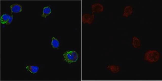
Immunohistochemistry Analysis: 2.5 µg/mL from a representative lot detected EPAC2 in mouse liver tissue.
Research Category
Neuroscience
Research Sub Category
Developmental Neuroscience

Quality

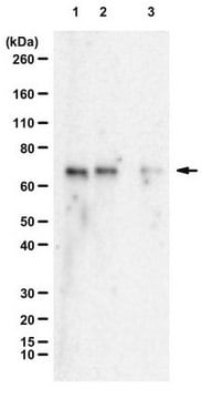
Evaluated by Western Blotting/Peptide Inhibition Assay in rat liver tissue lysate.

Western Blotting/Peptide Inhibition Assay Analysis: 1 µg/mL of this antibody detected EPAC2 in 15 µg of rat liver tissue lysate.

Target description

~129 kDa observed. Uncharacterized bands may appear in some lysate(s). Uniprot describes 3 isoforms at ~79 kDa, ~100 kDa, and ~116 kDa

Physical form

Antigen Affinity Purified
Purified rabbit polyclonal in buffer containing PBS with up to 0.1% sodium azide.

Storage and Stability

Stable for 1 year at 2-8°C from date of receipt.

Analysis Note

Control
Rat liver tissue lysate

Other Notes

Concentration: Please refer to the Certificate of Analysis for the lot-specific concentration.

Disclaimer

Unless otherwise stated in our catalog or other company documentation accompanying the product(s), our products are intended for research use only and are not to be used for any other purpose, which includes but is not limited to, unauthorized commercial uses, in vitro diagnostic uses, ex vivo or in vivo therapeutic uses or any type of consumption or application to humans or animals.

Not finding the right product?  

Try our Product Selector Tool.

Storage Class Code

10 - Combustible liquids

WGK

WGK 2

Flash Point(F)

Not applicable

Flash Point(C)

Not applicable


Regulatory Listings

Regulatory Listings are mainly provided for chemical products. Only limited information can be provided here for non-chemical products. No entry means none of the components are listed. It is the user’s obligation to ensure the safe and legal use of the product.

JAN Code

ABN492:


Certificates of Analysis (COA)

Search for Certificates of Analysis (COA) by entering the products Lot/Batch Number. Lot and Batch Numbers can be found on a product’s label following the words ‘Lot’ or ‘Batch’.

Already Own This Product?

Find documentation for the products that you have recently purchased in the Document Library.

Visit the Document Library

Michel Rasmussen et al.
Journal of neurochemistry, 157(6), 2173-2186 (2020-11-25)
The hereditary disease Retinitis pigmentosa results in severe vision loss due to photoreceptor degeneration by unclear mechanisms. In several disease models, the second messenger cGMP accumulates in the degenerating photoreceptors, where it may over-activate specific cGMP-interacting proteins, like cGMP-dependent protein
Michel Rasmussen et al.
International journal of molecular sciences, 23(9) (2022-05-15)
The disease retinitis pigmentosa (RP) leads to photoreceptor degeneration by a yet undefined mechanism(s). In several RP mouse models (i.e., rd mice), a high cyclic GMP (cGMP) level within photoreceptors is detected, suggesting that cGMP plays a role in degeneration.

Our team of scientists has experience in all areas of research including Life Science, Material Science, Chemical Synthesis, Chromatography, Analytical and many others.

Contact Technical Service