Skip to Content
Merck
All Photos(1)

Key Documents

350R-1

Sigma-Aldrich

FoxP1 (SP133) Rabbit Monoclonal Antibody

Sign Into View Organizational & Contract Pricing


About This Item

UNSPSC Code:
12352203
NACRES:
NA.41

biological source

rabbit

Quality Level

100
500

conjugate

unconjugated

antibody form

culture supernatant

antibody product type

primary antibodies

clone

SP133, monoclonal

description

For In Vitro Diagnostic Use in Select Regions (See Chart)

form

buffered aqueous solution

species reactivity

human

packaging

vial of 0.1 mL concentrate (350R-14)
vial of 0.5 mL concentrate (350R-15)
bottle of 1.0 mL predilute (350R-17)
vial of 1.0 mL concentrate (350R-16)
bottle of 7.0 mL predilute (350R-18)

manufacturer/tradename

Cell Marque

technique(s)

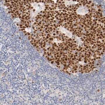
immunohistochemistry (formalin-fixed, paraffin-embedded sections): 1:100-1:500

isotype

IgG

control

DLBCL, lymph node, tonsil

shipped in

wet ice

storage temp.

2-8°C

visualization

nuclear

Gene Information

human ... FOXP1(27086)

General description

Diffuse large B-cell lymphoma (DLBCL) represents different clinicopathologic entities which are difficult to separate using standard techniques. From the clinical standpoint, the introduction of immunochemotherapy in the treatment of DLBCL has dramatically improved the outcome of these patients compared with chemotherapy alone. Gene expression profiling (GEP) studies have shown that DLBCL can be reproducibly divided into the important subtypes of germinal center B-cell–like (GCB), activated B-cell–like (ABC), and unclassified DLBCL. It is beneficial to translate the GEP classification into protein expression by tumor cells through immunohistochemical (IHC) staining of formalin-fixed, paraffin-embedded tissues. A panel of antibodies: CD10, BCL6, MUM1/IRF4, GCET1, FoxP1, LMO2, and BCL2 has been used to determine GCB or ABC and each has different percentage thresholds for positive staining. Choi et al. demonstrated that the cases positive for GCET1 (≥ 80% of tumor cells) and MUM1/IRF4 (≥ 80%) and/or FoxP1 (≥ 80%) or negative for CD10 and BCL6 (≤ 30%) were assigned to the group. The cases positive for CD10 (≥ 30%), GCET1 (≥ 80%) without MUM1 expression, or positive for BCL6 without FoxP1 expression were classified as GCB. This study indicated the importance of FoxP1 in the subclassification of DLBCL. Choi et al then modified their approach to DLBCL subclassification by focusing on FoxP1. The tumors that are positive for both FoxP1 and GCET1 are assigned to GCB subgroup, but, if FoxP1 is positive and GCET1 is negative, the tumors belong to the ABC phenotype. If a case is FoxP1 negative but MUM-1/IRF4 positive, it still belongs to the ABC phenotype as long as CD10 is not expressed. This modified method emphasized the role of FoxP1, MUM1/IRF4, and GCET1 in the subclassification of DLBCL. The Choi′s algorithm had a very high concordance with the GEP results (87%). Therefore, FoxP1 is useful in subclassification of DLBCL and a high cutoff (≥80%) for FoxP1 is needed to achieve high specificity for the ABC subtype.

Quality


IVD

IVD

IVD

RUO

Linkage

FoxP1 Positive Control Slides, Product No. 350S, are available for immunohistochemistry (formalin-fixed, paraffin-embedded sections).

Physical form

Solution in Tris Buffer, pH 7.3-7.7, with 1% BSA and <0.1% Sodium Azide

Preparation Note

Download the IFU specific to your product lot and formatNote: This requires a keycode which can be found on your packaging or product label.

Other Notes

For Technical Service please contact: 800-665-7284 or email: service@cellmarque.com

Legal Information

Cell Marque is a trademark of Merck KGaA, Darmstadt, Germany

Not finding the right product?  

Try our Product Selector Tool.


Certificates of Analysis (COA)

Search for Certificates of Analysis (COA) by entering the products Lot/Batch Number. Lot and Batch Numbers can be found on a product’s label following the words ‘Lot’ or ‘Batch’.

Already Own This Product?

Find documentation for the products that you have recently purchased in the Document Library.

Visit the Document Library

Paul N Meyer et al.
Journal of clinical oncology : official journal of the American Society of Clinical Oncology, 29(2), 200-207 (2010-12-08)
Patients with diffuse large B-cell lymphoma (DLBCL) can be divided into prognostic groups based on the cell of origin of the tumor as determined by microarray analysis. Various immunohistochemical algorithms have been developed to replicate these microarray results and/or stratify
Swerdlow, SH et al. WHO Classification of Tumours of Haematopoietic and Lymphoid Tissues. Geneva: World Health Organization; 2008.
WHO Classification of Tumours of Haematopoietic and Lymphoid Tissue (2008)
Swerdlow, SH et al. WHO Classification of Tumours of Haematopoietic and Lymphoid Tissues. Geneva: World Health Organization; 2008.
WHO Classification of Tumours of Haematopoietic and Lymphoid Tissue (2008)
H Hagberg et al.
Annals of oncology : official journal of the European Society for Medical Oncology, 17 Suppl 4, iv31-iv32 (2006-05-17)
The multicentre phase III CORAL study aims to guide choice of salvage chemotherapy in diffuse large B-cell lymphoma (DLBCL) and assess the role of rituximab maintenance after autologous stem cell transplantation (ASCT). Patients are first randomised between ICE (ifosfamide, carboplatin
The New England journal of medicine, 329(14), 987-994 (1993-09-30)
Although many patients with intermediate-grade or high-grade (aggressive) non-Hodgkin's lymphoma are cured by combination chemotherapy, the remainder are not cured and ultimately die of their disease. The Ann Arbor classification, used to determine the stage of this disease, does not

Our team of scientists has experience in all areas of research including Life Science, Material Science, Chemical Synthesis, Chromatography, Analytical and many others.

Contact Technical Service