Skip to Content
Merck
All Photos(5)

Key Documents

05-829

Sigma-Aldrich

Anti-α-Tubulin Antibody, clone DM1A

clone DM1A, Upstate®, from mouse

Synonym(s):

Alpha-tubulin 3, Tubulin B-alpha-1, Tubulin alpha-3 chain, tubulin, alpha 1a, tubulin, alpha 3, tubulin, alpha, brain-specific

Sign Into View Organizational & Contract Pricing


About This Item

UNSPSC Code:
12352203
eCl@ss:
32160702
NACRES:
NA.41

biological source

mouse

Quality Level

antibody form

purified immunoglobulin

clone

DM1A, monoclonal

species reactivity

avian, pig, rat, bovine, mouse, human, guinea pig

manufacturer/tradename

Upstate®

technique(s)

immunocytochemistry: suitable
immunofluorescence: suitable
western blot: suitable

isotype

IgG1

NCBI accession no.

UniProt accession no.

shipped in

dry ice

target post-translational modification

unmodified

Gene Information

human ... TUBA1A(7846)

General description

Microtubules are involved in a wide variety of cellular activities ranging from mitosis and transport events to cell movement and the maintenance of cell shape. Tubulin itself is a globular protein which consists of two polypeptides (alpha and beta tubulin). Alpha and beta tubulin dimers are assembled to 13 protofilaments that form a microtubule of 22 nm diameter. Tyrosine ligase ads a C-terminal tyrosin to monomeric alpha tubulin. Assembled microtubules can again be detyrosinated by a cytoskeleton associated carboxypeptidase. Detyrosinated alpha tubulin is referred to as Glu-tubulin. Another post-translational modification of detyrosinated alpha tubulin is C-terminal polyglutamylation which is characteristic for microtubules in neuronal cells and the mitotic spindle.

Specificity

Recognizes α-Tubulin, clone DM1A, MW 50-60 kDa. The epitope recognized by this antibody has been mapped to amino acids 426-450.

Immunogen

Epitope: a.a. 426-450
Purified human brain α-Tubulin. The epitope recognized by this antibody has been mapped to amino acids 426-450 (AALEKDYEEVGVDSVEGEGEEEGEE).

Application

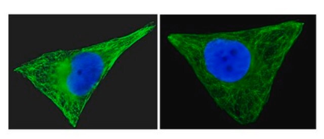
Anti-α-Tubulin Antibody, clone DM1A is an antibody against α-Tubulin for use in IC, IF & WB.
Immunofluorescence:
(Breitling, F., 1986)
Research Category
Cell Structure
Research Sub Category
Cytoskeleton

Quality

Routinely evaluated by western blot on non-stimulated A431 lysate.

Western Blot Analysis:
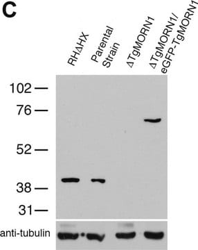
0.1-1 µg/mL of this lot detected α- Tubulin in RIPA lysates from A431, L6 cells and rat whole brain tissue.

Target description

50-60 kDa

Linkage

Replaces: 04-1117

Physical form

Format: Purified
Protein G Purified
Purified mouse monoclonal IgG1 in buffer containing 0.1M Tris-Glycine, 0.15M NaCl, 0.05% Sodium Azide and 30% glycerol.

Storage and Stability

Stable for 1 year at -20°C from date of receipt.

Handling Recommendations:
Upon first thaw, and prior to removing the cap, centrifuge the vial and gently mix the solution. Aliquot into microcentrifuge tubes and store at -20°C. Avoid repeated freeze/thaw cycles, which may damage IgG and affect product performance. Note: Variabillity in freezer temperatures below -20°C may cause glycerol containing solutions to become frozen during storage.

Analysis Note

Control
Positive Antigen Control: Catalog #12-301, non-stimulated A431 cell lysate. Add 2.5µL of 2-mercaptoethanol/100µL of lysate and boil for 5 minutes to reduce the preparation. Load 20µg of reduced lysate per lane for minigels.

Other Notes

Concentration: Please refer to the Certificate of Analysis for the lot-specific concentration.

Legal Information

UPSTATE is a registered trademark of Merck KGaA, Darmstadt, Germany

Disclaimer

Unless otherwise stated in our catalog or other company documentation accompanying the product(s), our products are intended for research use only and are not to be used for any other purpose, which includes but is not limited to, unauthorized commercial uses, in vitro diagnostic uses, ex vivo or in vivo therapeutic uses or any type of consumption or application to humans or animals.

Storage Class Code

10 - Combustible liquids

WGK

WGK 1


Certificates of Analysis (COA)

Search for Certificates of Analysis (COA) by entering the products Lot/Batch Number. Lot and Batch Numbers can be found on a product’s label following the words ‘Lot’ or ‘Batch’.

Already Own This Product?

Find documentation for the products that you have recently purchased in the Document Library.

Visit the Document Library

G protein binding and G protein activation by nucleotide transfer involve distinct domains on tubulin: regulation of signal transduction by cytoskeletal elements.
Roychowdhury, S, et al.
Biochemistry, 32, 4955-4961 (1993)
MicroRNA-7 inhibits multiple oncogenic pathways to suppress HER2?16 mediated breast tumorigenesis and reverse trastuzumab resistance.
Huynh, FC; Jones, FE
Testing null
H3 relaxin demonstrates antifibrotic properties via the RXFP1 receptor.
Hossain MA, Man BC, Zhao C, Xu Q, Du XJ, Wade JD, Samuel CS
Biochemistry null
Diana M Cittelly et al.
Carcinogenesis, 31(12), 2049-2057 (2010-09-30)
Tamoxifen is the most commonly prescribed therapy for patients with estrogen receptor (ER)α-positive breast tumors. Tumor resistance to tamoxifen remains a serious clinical problem especially in patients with tumors that also overexpress human epidermal growth factor receptor 2 (HER2). Current
Reactivation of epigenetically silenced HER4/ERBB4 results in apoptosis of breast tumor cells.
Das, PM; Thor, AD; Edgerton, SM; Barry, SK; Chen, DF; Jones, FE
Oncogene null

Our team of scientists has experience in all areas of research including Life Science, Material Science, Chemical Synthesis, Chromatography, Analytical and many others.

Contact Technical Service