General description
Ubiqutin (Ub) is initially produced as a 229-a.a. Polyubiquitin-B (UniProt P0CG47) precursor protein encoded by the UBB gene (Gene ID 7314) or a 685-a.a. Polyubiquitin-C precursor protein (UniProt P0CG48) encoded by the UBC gene (Gene ID 7316) in human. The C-termini of Polyubiquitin-B (Cys229) and Polyubiquitin-C (Val685) are post-translationally removed, followed by additional posttranslational cleavages to yield multiple copies of idential 76-amino acid ubiquitin (also known as high mobility group nonhistone protein HMG-20) molecules (three copies per Polyubiquitin-B and nine copies per Polyubiquitin-C). Covalent modifications of target proteins via different Ub Lys residues are well documented events during cellular DNA repair, ER-associated degradation (ERAD), transcription activation, lysosomal and proteasomal degradation. PINK1 (PTEN induced putative kinase 1) is a Ser/Thr kinase that specifically accumulates in depolarized mitochondria, where it acts as a positive regulator of parkin (Park2) E3 ubiquitin (Ub) ligase activity by phosphorylating Ub at Ser65 as well as the corresponding Ser residue in parkin N-terminal Ub-like (UBL) domain. Phosphorylated Ub interacts with phosphorylated parkin at its GINGO0 domain in an allosteric manner, inducing a conformation change and exposing parkin RING2 domain catalytic cysteine for full ligase activity. NMR based conformation study reveals that Ser65-phosphorylated Ub exists in two conformations in solution. The major conformation displays altered surface properties, while the second phosphoUb conformation exhibits a retracted C-terminal tail by two residues into the Ub core. Ub Ser65 phosphorylation has little effect toward E1-mediated E2 charging and mainly affects the discharging of E2 enzymes to form polyUb chains. In addition, the majority of deubiquitinases (DUBs), including USP8, USP15 and USP30, are shown to exhibit impaired activity against Lys63-linked poly-phosphoUb.
Specificity
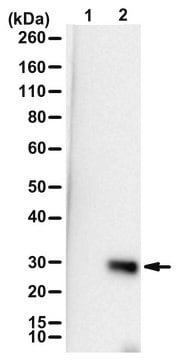
Target specificity was verified using the unconjugated antibody (ABS1513-I). Only Ser65-phosphorylated ubiquitin (Ub) and ubiquitinated proteins (including oligoUb and polyUb) containing Ser65-phosphorylated ubiquitin were detected. This antibody exhibits no reactivity toward Ub or ubiquitinated proteins in the absence of Ub Ser65 phosphorylation.
Immunogen
KLH-conjugated linear peptide corresponding to the ubiquitin target region sequence containing phosphorylated Ser65.
Application
Detect ubiquitin Ser65 phosphorylation using this rabbit polyclonal Anti-phospho-Ubiquitin (Ser65) Antibody, Alexa Fluor™ 488 Conjugate, Cat. No. ABS1513-I-AF488, validated for use in Immunocytochemistry.
Research Category
Signaling
The unconjugated antibody (Cat. No. AB1513-I) is also available for Immunocytochemistry and Western Blotting applications.
Quality
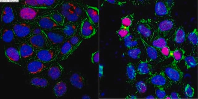
Evaluated by Immunocytochemistry in PINK1-expressing HeLa cells with or without CCCP treatment.
Immunocytochemistry Analysis: A 1:100 dilution of this antibody detected CCCP treatment-induced ubiquitin Ser65 phosphorylation in PINK1-expressing HeLa cells.
Target description
~17/8 kDa (dimer/monomer) and ~45-250 kDa observed. 8.565/17.112/25.66/34.21 kDa (mono-/di/tri-/tetra-ubiquitin) calculated. Bands ~45-250 kDa represent ubiquitinated proteins containing ser65-phosphorylated Ub. Uncharacterized bands may be observed in some lysate(s).
Physical form
Affinity purified.
Purified rabbit polyclonal antibody in PBS with 1.5% BSA and 0.05% sodium azide.
Storage and Stability
Stable for 1 year at 2-8°C from date of receipt.
Other Notes
Concentration: Please refer to lot specific datasheet.
Legal Information
ALEXA FLUOR is a trademark of Life Technologies
Disclaimer
Unless otherwise stated in our catalog or other company documentation accompanying the product(s), our products are intended for research use only and are not to be used for any other purpose, which includes but is not limited to, unauthorized commercial uses, in vitro diagnostic uses, ex vivo or in vivo therapeutic uses or any type of consumption or application to humans or animals.


