推荐产品
生物源
mouse
品質等級
抗體表格
purified immunoglobulin
抗體產品種類
primary antibodies
無性繁殖
8A6-F8, monoclonal
物種活性
rat, mouse, human
技術
immunohistochemistry: suitable (paraffin)
immunoprecipitation (IP): suitable
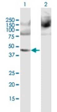
western blot: suitable
同型
IgG1κ
NCBI登錄號
UniProt登錄號
運輸包裝
ambient
目標翻譯後修改
unmodified
基因資訊
mouse ... Ppme1(72590)
一般說明
Protein phosphatase methylesterase 1 (UniProt: Q8BVQ5; also known as PME1; EC:3.1.1.89) is encoded by the Ppme1 (also known as Pme1) gene (Gene ID: 72590) in murine species. PME1 is a member of the AB hydrolase family that shows ubiquitous distribution, but is highly expressed in testis and brain. PME-1 is shown to be PMSF-resistant and okadaic acid-sensitive methyltransferase. PME-1 contains a motif found in lipases that utilize a catalytic triad-activated serine as their active site nucleophile, and has other scattered homology with other lipases in which this motif is conserved. PME1 catalyzes the demethylation of proteins that have been reversibly carboxymethylated. PME1 is shown to demethylate and inactivate protein phosphatase 2A (PP2A). Its binding to the catalytic site of PP2A displaces manganese ion that results in the inactivation of PP2A. Targeted disruption of the Ppme1 gene is shown to cause perinatal lethality in mice, resulting from a virtually complete loss of the demethylated form of PP2A in the nervous system and peripheral tissues.
特異性
Clone 8A6-F8 specifically stains PME-1 in paraffin sections of human pancreas, testis, and cerebral cortex.
免疫原
His-tagged full length recombinant mouse Protein phosphatase methylesterase 1.
應用
Detect Protein phosphatase methylesterase 1 using this mouse monoclonal Anti-PME1 Antibody, clone 8A6-F8, Cat. No. MABC1183. It is used in Immunohistochemistry (Paraffin), Immunoprecipitation, and Western Blotting.
Immunohistochemistry Analysis: A 1:50 dilution from a representative lot detected PME1 in human testis and human cerebral cortex tissue.
Immunoprecipitation Analysis: A representative lot detected PME1 in NIH3T3 mouse fibroblasts, wild-type, but not in PME1 knock-out mouse embryonic fibroblasts. (Courtesy of Stefan Schuchner, Ph.D. and Egon Ogris, M.D., Medical University of Vienna Austria).
Western Blotting Analysis: A 1:50 dilution from a representative lot detected PME1 in HeLa, NIH3T3, Rat1, BHK21, and CV-1 lysates. (Courtesy of Stefan Schuchner, Ph.D. and Egon Ogris, M.D., Medical University of Vienna Austria).
Research Category
Apoptosis & Cancer
Apoptosis & Cancer
品質
Evaluated by Immunohistochemistry in human pancreas tissue.
Immunohistochemistry Analysis: A 1:50 dilution of this antibody detected PME1 in human pancreas tissue.
Immunohistochemistry Analysis: A 1:50 dilution of this antibody detected PME1 in human pancreas tissue.
標靶描述
42.25 kDa calculated.
外觀
Protein G purified
Format: Purified
Purified mouse monoclonal antibody IgG1 in buffer containing 0.1 M Tris-Glycine (pH 7.4), 150 mM NaCl with 0.05% sodium azide.
儲存和穩定性
Stable for 1 year at 2-8°C from date of receipt.
其他說明
Concentration: Please refer to lot specific datasheet.
免責聲明
Unless otherwise stated in our catalog or other company documentation accompanying the product(s), our products are intended for research use only and are not to be used for any other purpose, which includes but is not limited to, unauthorized commercial uses, in vitro diagnostic uses, ex vivo or in vivo therapeutic uses or any type of consumption or application to humans or animals.
未找到合适的产品?
试试我们的产品选型工具.
儲存類別代碼
12 - Non Combustible Liquids
水污染物質分類(WGK)
WGK 1
Stefan Schüchner et al.
Scientific reports, 6, 31363-31363 (2016-08-18)
Western blotting is one of the most widely used techniques in molecular biology and biochemistry. Prestained proteins are used as molecular weight standards in protein electrophoresis. In the chemiluminescent Western blot analysis, however, these colored protein markers are invisible leaving
我们的科学家团队拥有各种研究领域经验,包括生命科学、材料科学、化学合成、色谱、分析及许多其他领域.
联系技术服务部门


