Skip to Content
Merck
All Photos(2)

Documents

P4124

Sigma-Aldrich

Anti-PM/Scl-100 antibody produced in rabbit

~1.0 mg/mL, affinity isolated antibody, buffered aqueous solution

Synonym(s):

Anti-Autoantigen PM/Scl 2, Anti-EXOSC10, Anti-Exsosome Component 10, Anti-P100, Anti-PMSCL Autoantigen, 100-KD, Anti-PMSCL2, Anti-Polymyositis-scleroderma Overlap Syndrome Associated Autoantigen, Anti-Polymyositis/scleroderma Autoantigen 2, Anti-Polymyositis/scleroderma Autoantigen, 100 kDa

Sign Into View Organizational & Contract Pricing


About This Item

MDL number:
UNSPSC Code:
12352203
NACRES:
NA.41

biological source

rabbit

conjugate

unconjugated

antibody form

affinity isolated antibody

antibody product type

primary antibodies

clone

polyclonal

form

buffered aqueous solution

mol wt

antigen ~100 kDa

species reactivity

human, rat, mouse

concentration

~1.0 mg/mL

technique(s)

indirect immunofluorescence: 1-2 μg/mL using rat NRK and mouse NIH3T3 cells
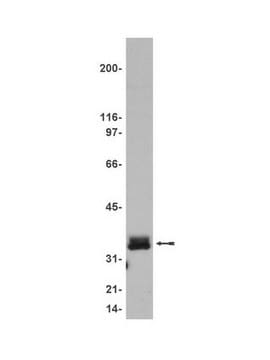
western blot (chemiluminescent): 1-2 μg/mL using nuclear exracts to HeLa human epithelioid carcinoma cells

UniProt accession no.

shipped in

dry ice

storage temp.

−20°C

target post-translational modification

unmodified

Gene Information

human ... EXOSC10(5394)
mouse ... Exosc10(50912)

General description

PM/SCL-100 (human exosome) is also known as RRP6, EXOSC10 (exosome component 10). It is a heterogeneous autoantibody that is located on human chromosome 1p36. In mouse, EXOSC10 is located on XY body.

Immunogen

synthetic peptide corresponding to amino acid residues 231-245 of human PM/Scl-100 with an N-terminal added cysteine, conjugated to KLH. The corresponding sequence is identical in both rat and mouse PM/Scl-100.

Application

Applications in which this antibody has been used successfully, and the associated peer-reviewed papers, are given below.
Immunofluorescence (1 paper)
Western Blotting (1 paper)

Biochem/physiol Actions

PM/SCL-100 (human exosome) participates in ribosome biogenesis, snoRNA (small nucleolar RNAs)processing, surveillance and degradation of nonfunctional transcripts. It is essential for the production of 40S subunit and to maintain global translation. EXOSC10 participates in mitotic cell division. In mouse, Exosc10 is required for spermatogonial proliferation and early embryogenesis.

Physical form

Solution in 0.01 M phosphate buffered saline, pH 7.4, and 15 mM sodium azide.

Disclaimer

Unless otherwise stated in our catalog or other company documentation accompanying the product(s), our products are intended for research use only and are not to be used for any other purpose, which includes but is not limited to, unauthorized commercial uses, in vitro diagnostic uses, ex vivo or in vivo therapeutic uses or any type of consumption or application to humans or animals.

Not finding the right product?  

Try our Product Selector Tool.

Storage Class Code

10 - Combustible liquids

WGK

WGK 3

Flash Point(F)

Not applicable

Flash Point(C)

Not applicable


Certificates of Analysis (COA)

Search for Certificates of Analysis (COA) by entering the products Lot/Batch Number. Lot and Batch Numbers can be found on a product’s label following the words ‘Lot’ or ‘Batch’.

Already Own This Product?

Find documentation for the products that you have recently purchased in the Document Library.

Visit the Document Library

Cooling-induced SUMOylation of EXOSC10 down-regulates ribosome biogenesis.
Knight JR et al
RNA, 22(4), 623-635 (2016)
Antibodies against PM/Scl-75 and PM/Scl-100 are independent markers for different subsets of systemic sclerosis patients.
Hanke K et al
Arthritis Research & Therapy, 11(1), R22-R22 (2009)
EXOSC10/Rrp6 is post-translationally regulated in male germ cells and controls the onset of spermatogenesis.
Jamin SP, et al.
Scientific Reports, 7(1), 15065-15065 (2017)
Activities of human RRP6 and structure of the human RRP6 catalytic domain.
Januszyk K, et al.
RNA, 17(8), 1566-1577 (2011)
Differential expression between human dermal papilla cells from balding and non-balding scalps reveals new candidate genes for androgenetic alopecia.
Chew EG, et al.
The Journal of Investigative Dermatology, 136(8), 1559-1567 (2016)

Our team of scientists has experience in all areas of research including Life Science, Material Science, Chemical Synthesis, Chromatography, Analytical and many others.

Contact Technical Service